 ムービー
ムービー コントローラー
 構造ビューア
 EMN検索について


-検索条件
-検索結果
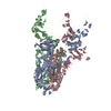
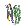
検索 (著者・登録者: du & m)の結果4,162件中、1から50件目までを表示しています

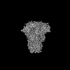
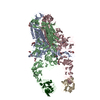
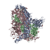
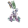
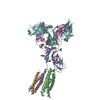
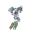
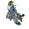
EMDB-39025: 
Structure of HCoV-HKU1A spike in the functionally anchored-3up conformation with 3TMPRSS2

EMDB-39026:
Local structure of HCoV-HKU1A spike in complex with TMPRSS2 and glycan

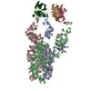
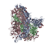
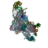
EMDB-39036:
Structure of HCoV-HKU1C spike in the functionally anchored-1up conformation with 1TMPRSS2
EMDB-39037:
Structure of HCoV-HKU1C spike in the functionally anchored-2up conformation with 2TMPRSS2
EMDB-39038:
Structure of HCoV-HKU1C spike in the functionally anchored-3up conformation with 2TMPRSS2

EMDB-39039:
Structure of HCoV-HKU1C spike in the functionally anchored-3up conformation with 3TMPRSS2

EMDB-39040:
Local structure of HCoV-HKU1C spike in complex with TMPRSS2 and glycan
EMDB-39041:
Structure of HCoV-HKU1C spike in the inactive-closed conformation
EMDB-39042:
Structure of HCoV-HKU1C spike in the inactive-1up conformation

EMDB-39043:
Structure of HCoV-HKU1C spike in the inactive-2up conformation
EMDB-39044:
Structure of HCoV-HKU1C spike in the glycan-activated-closed conformation

EMDB-39045:
Structure of HCoV-HKU1C spike in the glycan-activated-1up conformation

EMDB-39046:
Structure of HCoV-HKU1C spike in the glycan-activated-2up conformation

EMDB-39047:
Structure of HCoV-HKU1C spike in the glycan-activated-3up conformation

EMDB-39048:
Local structure of HCoV-HKU1C spike in complex with glycan
PDB-8y7x:
Structure of HCoV-HKU1A spike in the functionally anchored-3up conformation with 3TMPRSS2
PDB-8y7y:
Local structure of HCoV-HKU1A spike in complex with TMPRSS2 and glycan
PDB-8y87:
Structure of HCoV-HKU1C spike in the functionally anchored-1up conformation with 1TMPRSS2
PDB-8y88:
Structure of HCoV-HKU1C spike in the functionally anchored-2up conformation with 2TMPRSS2
PDB-8y89:
Structure of HCoV-HKU1C spike in the functionally anchored-3up conformation with 2TMPRSS2
PDB-8y8a:
Structure of HCoV-HKU1C spike in the functionally anchored-3up conformation with 3TMPRSS2
PDB-8y8b:
Local structure of HCoV-HKU1C spike in complex with TMPRSS2 and glycan
PDB-8y8c:
Structure of HCoV-HKU1C spike in the inactive-closed conformation

PDB-8y8d:
Structure of HCoV-HKU1C spike in the inactive-1up conformation
PDB-8y8e:
Structure of HCoV-HKU1C spike in the inactive-2up conformation
PDB-8y8f:
Structure of HCoV-HKU1C spike in the glycan-activated-closed conformation

PDB-8y8g:
Structure of HCoV-HKU1C spike in the glycan-activated-1up conformation
PDB-8y8h:
Structure of HCoV-HKU1C spike in the glycan-activated-2up conformation
PDB-8y8i:
Structure of HCoV-HKU1C spike in the glycan-activated-3up conformation
PDB-8y8j:
Local structure of HCoV-HKU1C spike in complex with glycan
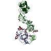
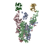
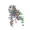
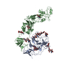
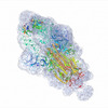
EMDB-39033:
Structure of the human ige-fc bound to its high affinity receptor fc(epsilon)
PDB-8z0t:
Structure of the human ige-fc bound to its high affinity receptor fc(epsilon)

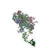
EMDB-60089:
Structure of the ige-fc bound to its high affinity receptor fc(epsilon)ri state2

EMDB-60090:
Structure of the ige-fc bound to its high affinity receptor fc(epsilon)ri state3
PDB-8zgs:
Structure of the ige-fc bound to its high affinity receptor fc(epsilon)ri state2
PDB-8zgt:
Structure of the ige-fc bound to its high affinity receptor fc(epsilon)ri state3
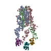
EMDB-39029:
Structure of the ige-fc bound to its high affinity receptor fc(epsilon)ri
PDB-8y81:
Structure of the ige-fc bound to its high affinity receptor fc(epsilon)ri

EMDB-39032:
Structure of the high affinity receptor fc(epsilon)ri TM
PDB-8y84:
Structure of the high affinity receptor fc(epsilon)ri TM
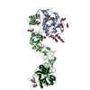
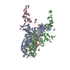
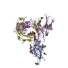
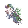
EMDB-17295:
Stabilised BA.1 SARS-CoV-2 spike with H6 nanobodies in '3 up' RBD conformation
PDB-8oyt:
Stabilised BA.1 SARS-CoV-2 spike with H6 nanobodies in '3 up' RBD conformation
EMDB-18438:
mt-SSU assembly intermediate in GTPBP8 knock-out cells, state 1

EMDB-18439:
mt-SSU assembly intermediate in GTPBP8 knock-out cells, state 2

EMDB-18440:
mt-SSU assembly intermediate in GTPBP8 knock-out cells, state 3

EMDB-18443:
mt-SSU assembly intermediate in GTPBP8 knock-out cells, state 4

EMDB-18460:
mt-LSU assembly intermediate in GTPBP8 knock-out cells, state 1

EMDB-18461:
mt-LSU assembly intermediate in GTPBP8 knock-out cells, state 2
PDB-8qrk:
mt-SSU assembly intermediate in GTPBP8 knock-out cells, state 1
PDB-8qrl:
mt-SSU assembly intermediate in GTPBP8 knock-out cells, state 2
ページ:
 wwPDBはEMDBデータモデルのバージョン3へ移行します
