 Movie
Movie Controller

[English] 日本語
 Yorodumi
- PDB-8z0t: Structure of the human ige-fc bound to its high affinity receptor... -
+ Open data

- Basic information
| Entry | Database: PDB / ID: 8z0t | ||||||
|---|---|---|---|---|---|---|---|
| Title | Structure of the human ige-fc bound to its high affinity receptor fc(epsilon) | ||||||
 Components Components |
| ||||||
 Keywords Keywords | ANTIMICROBIAL PROTEIN / COMPLEX | ||||||
| Function / homology |  Function and homology information Function and homology informationhigh-affinity IgE receptor activity / mononuclear cell differentiation / IgE B cell receptor complex / adaptive immune memory response / primary adaptive immune response / B cell antigen processing and presentation / type I hypersensitivity / Fc receptor-mediated immune complex endocytosis / eosinophil degranulation / IgE immunoglobulin complex ...high-affinity IgE receptor activity / mononuclear cell differentiation / IgE B cell receptor complex / adaptive immune memory response / primary adaptive immune response / B cell antigen processing and presentation / type I hypersensitivity / Fc receptor-mediated immune complex endocytosis / eosinophil degranulation / IgE immunoglobulin complex / macrophage activation / IgE binding / antibody-dependent cellular cytotoxicity / Fc epsilon receptor (FCERI) signaling / type 2 immune response / immunoglobulin complex, circulating / immunoglobulin receptor binding / mast cell degranulation / immunoglobulin mediated immune response / B cell proliferation / B cell activation / macrophage differentiation / Role of LAT2/NTAL/LAB on calcium mobilization / complement activation, classical pathway / antigen binding / FCERI mediated Ca+2 mobilization / B cell receptor signaling pathway / FCERI mediated MAPK activation / FCERI mediated NF-kB activation / antibacterial humoral response / Interleukin-4 and Interleukin-13 signaling / adaptive immune response / cell surface receptor signaling pathway / immune response / inflammatory response / external side of plasma membrane / cell surface / : / extracellular region / plasma membrane Similarity search - Function | ||||||
| Biological species |  Homo sapiens (human) Homo sapiens (human) | ||||||
| Method | ELECTRON MICROSCOPY / single particle reconstruction / cryo EM / Resolution: 3.58 Å | ||||||
 Authors Authors | Du, S. / Deng, M.J. / Xiao, J.Y. | ||||||
| Funding support | 1items
| ||||||
 Citation Citation | Journal: Nature / Year: 2024 Title: Structural insights into the high-affinity IgE receptor FcεRI complex. Authors: Meijie Deng / Shuo Du / Handi Hou / Junyu Xiao /  Abstract: Immunoglobulin E (IgE) plays a pivotal role in allergic responses. The high-affinity IgE receptor, FcεRI, found on mast cells and basophils, is central to the effector functions of IgE. FcεRI is a ...Immunoglobulin E (IgE) plays a pivotal role in allergic responses. The high-affinity IgE receptor, FcεRI, found on mast cells and basophils, is central to the effector functions of IgE. FcεRI is a tetrameric complex, comprising FcεRIα, FcεRIβ and a homodimer of FcRγ (originally known as FcεRIγ), with FcεRIα recognizing the Fc region of IgE (Fcε) and FcεRIβ-FcRγ facilitating signal transduction. Additionally, FcRγ is a crucial component of other immunoglobulin receptors, including those for IgG (FcγRI and FcγRIIIA) and IgA (FcαRI). However, the molecular basis of FcεRI assembly and the structure of FcRγ have remained elusive. Here we elucidate the cryogenic electron microscopy structure of the Fcε-FcεRI complex. FcεRIα has an essential role in the receptor's assembly, interacting with FcεRIβ and both FcRγ subunits. FcεRIβ is structured as a compact four-helix bundle, similar to the B cell antigen CD20. The FcRγ dimer exhibits an asymmetric architecture, and coils with the transmembrane region of FcεRIα to form a three-helix bundle. A cholesterol-like molecule enhances the interaction between FcεRIβ and the FcεRIα-FcRγ complex. Our mutagenesis analyses further indicate similarities between the interaction of FcRγ with FcεRIα and FcγRIIIA, but differences in that with FcαRI. These findings deepen our understanding of the signalling mechanisms of FcεRI and offer insights into the functionality of other immune receptors dependent on FcRγ. | ||||||
| History |
|
- Structure visualization
| Structure viewer | Molecule: MolmilJmol/JSmol |
|---|
- Downloads & links
-Download
| PDBx/mmCIF format | 8z0t.cif.gz | 160.5 KB | Display | PDBx/mmCIF format |
|---|---|---|---|---|
| PDB format | pdb8z0t.ent.gz | 124.6 KB | Display | PDB format |
| PDBx/mmJSON format | 8z0t.json.gz | Tree view | PDBx/mmJSON format | |
| Others |  Other downloads Other downloads |
-Validation report
| Arichive directory | https://data.pdbj.org/pub/pdb/validation_reports/z0/8z0tftp://data.pdbj.org/pub/pdb/validation_reports/z0/8z0t | HTTPS FTP |
|---|
-Related structure data
| Related structure data | 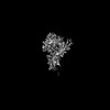 39033MC 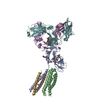 8y81C 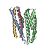 8y84C 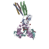 8zgsC 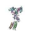 8zgtC M: map data used to model this data C: citing same article ( |
|---|---|
| Similar structure data |


-Links
 PDBj
PDBj


- Assembly
| Deposited unit | 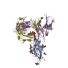
|
|---|---|
| 1 |
|
-Components
-Protein , 2 types, 3 molecules ADE
| #1: Protein | Mass: 29624.746 Da / Num. of mol.: 1 Source method: isolated from a genetically manipulated source Source: (gene. exp.) Homo sapiens (human) / Gene: FCER1A, FCE1A / Cell line (production host): HEK293 / Production host: Homo sapiens (human) / References: UniProt: P12319 |
|---|---|
| #2: Protein | Mass: 39368.285 Da / Num. of mol.: 2 Source method: isolated from a genetically manipulated source Source: (gene. exp.) Homo sapiens (human) / Gene: IGHE / Cell line (production host): HEK293 / Production host: Homo sapiens (human) / References: UniProt: P01854 |
-Sugars , 4 types, 9 molecules 
| #3: Polysaccharide | 2-acetamido-2-deoxy-beta-D-glucopyranose-(1-4)-beta-D-mannopyranose-(1-4)-2-acetamido-2-deoxy-beta- ...2-acetamido-2-deoxy-beta-D-glucopyranose-(1-4)-beta-D-mannopyranose-(1-4)-2-acetamido-2-deoxy-beta-D-glucopyranose-(1-4)-2-acetamido-2-deoxy-beta-D-glucopyranose Source method: isolated from a genetically manipulated source | ||
|---|---|---|---|
| #4: Polysaccharide | 2-acetamido-2-deoxy-beta-D-glucopyranose-(1-4)-2-acetamido-2-deoxy-beta-D-glucopyranose Source method: isolated from a genetically manipulated source | ||
| #5: Polysaccharide | Source method: isolated from a genetically manipulated source #6: Sugar | ChemComp-NAG /  Type: D-saccharide, beta linking / Mass: 221.208 Da / Num. of mol.: 5 / Source method: obtained synthetically / Formula: C8H15NO6 / Feature type: SUBJECT OF INVESTIGATION Type: D-saccharide, beta linking / Mass: 221.208 Da / Num. of mol.: 5 / Source method: obtained synthetically / Formula: C8H15NO6 / Feature type: SUBJECT OF INVESTIGATION |
-Details
| Has ligand of interest | Y |
|---|---|
| Has protein modification | Y |
-Experimental details
-Experiment
| Experiment | Method: ELECTRON MICROSCOPY |
|---|---|
| EM experiment | Aggregation state: PARTICLE / 3D reconstruction method: single particle reconstruction |
- Sample preparation
| Component | Name: the human ige-fc bound to its high affinity receptor fc(epsilon) Type: COMPLEX / Entity ID: #1-#2 / Source: RECOMBINANT |
|---|---|
| Source (natural) | Organism: Homo sapiens (human) |
| Source (recombinant) | Organism: Homo sapiens (human) |
| Buffer solution | pH: 7.4 |
| Specimen | Embedding applied: NO / Shadowing applied: NO / Staining applied: NO / Vitrification applied: YES |
| Vitrification | Cryogen name: ETHANE |
- Electron microscopy imaging
| Experimental equipment |  Model: Titan Krios / Image courtesy: FEI Company |
|---|---|
| Microscopy | Model: FEI TITAN KRIOS |
| Electron gun | Electron source:  FIELD EMISSION GUN / Accelerating voltage: 300 kV / Illumination mode: FLOOD BEAM FIELD EMISSION GUN / Accelerating voltage: 300 kV / Illumination mode: FLOOD BEAM |
| Electron lens | Mode: BRIGHT FIELD / Nominal defocus max: 1500 nm / Nominal defocus min: 1000 nm |
| Image recording | Electron dose: 60 e/Å2 / Film or detector model: GATAN K3 (6k x 4k) |
- Processing
| EM software | Name: PHENIX / Category: model refinement | ||||||||||||||||||||||||
|---|---|---|---|---|---|---|---|---|---|---|---|---|---|---|---|---|---|---|---|---|---|---|---|---|---|
| CTF correction | Type: PHASE FLIPPING AND AMPLITUDE CORRECTION | ||||||||||||||||||||||||
| 3D reconstruction | Resolution: 3.58 Å / Resolution method: FSC 0.143 CUT-OFF / Num. of particles: 101369 / Symmetry type: POINT | ||||||||||||||||||||||||
| Refine LS restraints |
|
