 Movie
Movie Controller
 Structure viewers
 About EMN search


-Search query
-Search result
Showing 1 - 50 of 53 items for (author: darrow & mc)

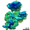
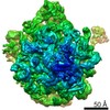
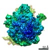
EMDB-51917: 
Streptavidin map obtained from graphene oxide modified self-wicking grids
Method: single particle / : Weckener M, Darrow MC, Clare DK, Naismith JH

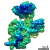
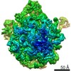
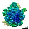
EMDB-51440:
3D reconstruction of beta-galactosidase obtained from self-wicking grids
Method: single particle / : Weckener M, Clare DK, Darrow MC, Naismith JH

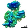
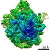
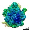
EMDB-51415:
3D reconstruction of beta-galactosidase obtained from graphene oxide modified self-wicking grids
Method: single particle / : Weckener M, Clare DK, Darrow MC, Naismith JH

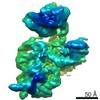
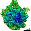
EMDB-51920:
KtrA.ADP with a 54ms plunge time on the chameleon.
Method: single particle / : Hirst IJ, Muench SP, Darrow MC, Scarff CA, Thompson RF

EMDB-51919:
Grid prepared using the vitrobot of KtrA.ADP from B. subtilis, small dataset
Method: single particle / : Hirst IJ, Muench SP

EMDB-51921:
KtrA.ADP with a 100ms plunge time on the chameleon
Method: single particle / : Hirst IJ, Muench SP

EMDB-51922:
KtrA.ADP with a 300ms plunge time on the chameleon
Method: single particle / : Hirst IJ, Muench SP, Darrow MC, Scarff CA, Thompson RF

EMDB-51923:
KtrA.ADP with 2500ms plunge time on the chameleon
Method: single particle / : Hirst IJ, Muench SP, Darrow MC, Scarff CA, Thompson RF

EMDB-51924:
KtrA.ADP with cyclic di-AMP prepared on the vitrobot
Method: single particle / : Hirst IJ, Muench SP, Darrow MC, Scarff CA, Thompson RF

EMDB-51925:
KtrA.ADP, chameleon with 180ms plunge time and DDM added
Method: single particle / : Hirst IJ, Muench SP, Darrow MC, Scarff CA, Thompson RF

EMDB-51926:
KtrA.ADP from chameleon with 2500ms plunge time and DDM added as a surfactant
Method: single particle / : Hirst IJ, Muench SP, Darrow MC, Scarff CA, Thompson RF

EMDB-51927:
KtrA.ADP with cyclic di-AMP from chameleon with 100ms plunge time.
Method: single particle / : Hirst IJ, Muench SP, Darrow MC, Scarff CA, Thompson RF

EMDB-51928:
KtrA.ADP prepared on the vitrobot, full particle stack
Method: single particle / : Hirst IJ, Muench SP

EMDB-15944:
CryoEM structure of GroEL-GroES-ADP.AlF3-Rubisco, class II.
Method: single particle / : Gardner S, Saibil HR
EMDB-15940:
CryoEM structure of GroEL-ADP.BeF3-Rubisco.
Method: single particle / : Gardner S, Saibil HR
EMDB-15942:
CryoEM structure of GroEL-GroES-ADP.AlF3-Rubisco.
Method: single particle / : Gardner S, Saibil HR
EMDB-15939:
CryoEM structure of nucleotide-free GroEL-Rubisco.
Method: single particle / : Gardner S, Saibil HR

EMDB-15941:
CryoEM reconstruction of GroEL-ATP-Rubisco.
Method: single particle / : Gardner S, Saibil HR
EMDB-15943:
CryoEM reconstruction of GroEL-GroES-ADP.AlF3-Rubisco, class I.
Method: single particle / : Gardner S, Saibil HR
EMDB-15945:
CryoEM reconstruction of GroEL-GroES-ADP.AlF3-Rubisco, class III.
Method: single particle / : Gardner S, Saibil HR
EMDB-15946:
CryoEM reconstruction of GroEL-GroES-ADP.AlF3-Rubisco, class IV.
Method: single particle / : Gardner S, Saibil HR

EMDB-17767:
Cryo electron tomography of human choriocarcinoma cells
Method: electron tomography / : Tun WM, Yee NB-Y, Ho EML, Darrow MC, Basham M
EMDB-15636:
Human 80S ribosome structure from pFIB-lamellae
Method: subtomogram averaging / : Berger C, Grange M

EMDB-16185:
80S human ribosome structure from PFIB lamellae of HeLa cells for assessing the extend and depth of the damage layer: 15 to 30 nm
Method: subtomogram averaging / : Berger C, Grange M

EMDB-16186:
80S human ribosome structure from PFIB lamellae of HeLa cells for assessing the extend and depth of the damage layer: above 30 nm matched control (for 15 to 30 nm)
Method: subtomogram averaging / : Berger C, Grange M
EMDB-16192:
80S human ribosome structure from PFIB lamellae of HeLa cells for assessing the extend and depth of the damage layer:30 to 45 nm
Method: subtomogram averaging / : Berger C, Grange M

EMDB-16193:
80S human ribosome structure from PFIB lamellae of HeLa cells for assessing the extend and depth of the damage layer: above 45 nm matched control (for 30 to 45 nm)
Method: subtomogram averaging / : Berger C, Grange M

EMDB-16194:
80S human ribosome structure from PFIB lamellae of HeLa cells for assessing the extend and depth of the damage layer:45 to 60 nm
Method: subtomogram averaging / : Berger C, Grange M

EMDB-16195:
80S human ribosome structure from PFIB lamellae of HeLa cells for assessing the extend and depth of the damage layer: above 60 nm matched control (for 45 to 60 nm)
Method: subtomogram averaging / : Berger C, Grange M

EMDB-16196:
80S human ribosome structure from PFIB lamellae of HeLa cells for assessing the extend and depth of the damage layer: 0 to 15 nm
Method: subtomogram averaging / : Berger C, Grange M

EMDB-16199:
80S human ribosome structure from PFIB lamellae of HeLa cells for assessing the extend and depth of the damage layer: above 15 nm matched control (for 0 to 15 nm)
Method: subtomogram averaging / : Berger C, Grange M

EMDB-23773:
Structure of the Neisseria gonorrhoeae ribonucleotide reductase in the inactive state
Method: single particle / : Levitz TS, Drennan CL
EMDB-10871:
30S ribosome subunit deposited by spraying (13 ms delay)
Method: single particle / : Klebl DP, Gravett MSC, Darrow M, Thompson RF, Muench SP
EMDB-10872:
30S ribosome subunit deposited using the chameleon (54 ms delay)
Method: single particle / : Klebl DP, Gravett MSC, Darrow M, Thompson RF, Muench SP
EMDB-10873:
30S ribosome subunit deposited using the chameleon (200 ms delay)
Method: single particle / : Klebl DP, Gravett MSC, Darrow M, Thompson RF, Muench SP
EMDB-10874:
30S ribosome subunit prepared by blotting
Method: single particle / : Klebl DP, Gravett MSC, Darrow M, Thompson RF, Muench SP
EMDB-10875:
50S ribosome subunit deposited by spraying (13 ms delay)
Method: single particle / : Klebl DP, Gravett MSC, Darrow M, Thompson RF, Muench SP
EMDB-10876:
50S ribosome subunit deposited using the chameleon (54 ms delay)
Method: single particle / : Klebl DP, Gravett MSC, Darrow M, Thompson RF, Muench SP
EMDB-10877:
50S ribosome subunit deposited using the chameleon (200 ms delay)
Method: single particle / : Klebl DP, Gravett MSC, Darrow M, Thompson RF, Muench SP
EMDB-10878:
50S ribosome subunit prepared by blotting
Method: single particle / : Klebl DP, Gravett MSC, Darrow M, Thompson RF, Muench SP
EMDB-10879:
70S ribosome deposited by spraying (13 ms delay)
Method: single particle / : Klebl DP, Gravett MSC, Darrow M, Thompson RF, Muench SP

EMDB-10880:
70S ribosome deposited using the chameleon (54 ms delay)
Method: single particle / : Klebl DP, Gravett MSC, Darrow M, Thompson RF, Muench SP
EMDB-10881:
70S ribosome deposited using the chameleon (200 ms delay)
Method: single particle / : Klebl DP, Gravett MSC, Darrow M, Thompson RF, Muench SP
EMDB-10882:
70S ribosome prepared by blotting
Method: single particle / : Klebl DP, Gravett MSC, Darrow M, Thompson RF, Muench SP

EMDB-10883:
HSPD1 single ring deposited by spraying (6 ms delay)
Method: single particle / : Klebl DP, Gravett MSC, Darrow M, Thompson RF, Muench SP

EMDB-10884:
HSPD1 single ring deposited by spraying (50 ms delay)
Method: single particle / : Klebl DP, Gravett MSC, Darrow M, Thompson RF, Muench SP

EMDB-10885:
HSPD1 single ring deposited using the chameleon (54 ms delay)
Method: single particle / : Klebl DP, Gravett MSC, Darrow M, Thompson RF, Muench SP

EMDB-10886:
HSPD1 single ring prepared by blotting
Method: single particle / : Klebl DP, Gravett MSC, Darrow M, Thompson RF, Muench SP

EMDB-8173:
CryoET of MEF Cells and Associated Manual Segmentation of Microtubule and Actin
Method: electron tomography / : Hecksel CW, Darrow MC, Dai W, Galaz-Montoya JG, Chin JA, Mitchell PG, Chen S, Jakana J, Schmid MF, Chiu W
EMDB-8174:
CryoET of U2OS Cells and Associated Manual Segmentation of Mitochondria
Method: electron tomography / : Hecksel CW, Darrow MC, Dai W, Galaz-Montoya JG, Chin JA, Mitchell PG, Chen S, Jakana J, Schmid MF, Chiu W
Pages:
 wwPDB to switch to version 3 of the EMDB data model
