Movie
Movie Controller


+ Open data

- Basic information
| Entry | Database: PDB / ID: 5nej | ||||||||||||
|---|---|---|---|---|---|---|---|---|---|---|---|---|---|
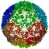
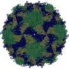
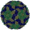
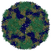
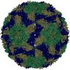
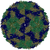
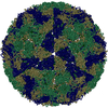
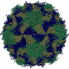
| Title | CryoEM Structure of Foot and Mouth Disease Virus O1 Manisa | ||||||||||||
 Components Components |
| ||||||||||||
 Keywords Keywords |  VIRUS / Foot and Mouth Disease Virus / FMDV / OpanAsia VIRUS / Foot and Mouth Disease Virus / FMDV / OpanAsia | ||||||||||||
| Function / homology |  Function and homology information Function and homology informationicosahedral viral capsid / modulation by virus of host chromatin organization / RNA-protein covalent cross-linking / ribonucleoside triphosphate phosphatase activity / T=pseudo3 icosahedral viral capsid / host cell cytoplasmic vesicle membrane / cytoplasmic vesicle membrane / : / regulation of translation / protein complex oligomerization ...icosahedral viral capsid / modulation by virus of host chromatin organization / RNA-protein covalent cross-linking / ribonucleoside triphosphate phosphatase activity / T=pseudo3 icosahedral viral capsid / host cell cytoplasmic vesicle membrane / cytoplasmic vesicle membrane / : / regulation of translation / protein complex oligomerization / monoatomic ion channel activity / clathrin-dependent endocytosis of virus by host cell / host cell cytoplasm / RNA helicase activity / viral protein processing / symbiont entry into host cell / viral RNA genome replication / cysteine-type endopeptidase activity / RNA-dependent RNA polymerase activity / DNA-templated transcription / virion attachment to host cell / structural molecule activity / proteolysis / RNA binding / ATP binding / cytoplasmSimilarity search - Function | ||||||||||||
| Biological species |  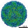 Foot-and-mouth disease virus Foot-and-mouth disease virus | ||||||||||||
| Method | ELECTRON MICROSCOPY / single particle reconstruction / cryo EM / Resolution: 3.1 Å | ||||||||||||
 Authors Authors | Kotecha, A. / Stuart, D. | ||||||||||||
| Funding support |  United Kingdom, 3items United Kingdom, 3items
| ||||||||||||
 Citation Citation | Journal: Nat Commun / Year: 2017 Title: Rules of engagement between αvβ6 integrin and foot-and-mouth disease virus. Authors: Abhay Kotecha / Quan Wang / Xianchi Dong / Serban L Ilca / Marina Ondiviela / Rao Zihe / Julian Seago / Bryan Charleston / Elizabeth E Fry / Nicola G A Abrescia / Timothy A Springer / Juha T ...Authors: Abhay Kotecha / Quan Wang / Xianchi Dong / Serban L Ilca / Marina Ondiviela / Rao Zihe / Julian Seago / Bryan Charleston / Elizabeth E Fry / Nicola G A Abrescia / Timothy A Springer / Juha T Huiskonen / David I Stuart /    Abstract: Foot-and-mouth disease virus (FMDV) mediates cell entry by attachment to an integrin receptor, generally αvβ6, via a conserved arginine-glycine-aspartic acid (RGD) motif in the exposed, antigenic, ...Foot-and-mouth disease virus (FMDV) mediates cell entry by attachment to an integrin receptor, generally αvβ6, via a conserved arginine-glycine-aspartic acid (RGD) motif in the exposed, antigenic, GH loop of capsid protein VP1. Infection can also occur in tissue culture adapted virus in the absence of integrin via acquired basic mutations interacting with heparin sulphate (HS); this virus is attenuated in natural infections. HS interaction has been visualized at a conserved site in two serotypes suggesting a propensity for sulfated-sugar binding. Here we determined the interaction between αvβ6 and two tissue culture adapted FMDV strains by cryo-electron microscopy. In the preferred mode of engagement, the fully open form of the integrin, hitherto unseen at high resolution, attaches to an extended GH loop via interactions with the RGD motif plus downstream hydrophobic residues. In addition, an N-linked sugar of the integrin attaches to the previously identified HS binding site, suggesting a functional role. | ||||||||||||
| History |
|
- Structure visualization
| Movie |
Movie viewer |
|---|---|
| Structure viewer | Molecule: MolmilJmol/JSmol |
- Downloads & links
-Download
| PDBx/mmCIF format | 5nej.cif.gz | 123.9 KB | Display | PDBx/mmCIF format |
|---|---|---|---|---|
| PDB format | pdb5nej.ent.gz | 99.6 KB | Display | PDB format |
| PDBx/mmJSON format | 5nej.json.gz | Tree view | PDBx/mmJSON format | |
| Others |  Other downloads Other downloads |
-Validation report
| Arichive directory | https://data.pdbj.org/pub/pdb/validation_reports/ne/5nejftp://data.pdbj.org/pub/pdb/validation_reports/ne/5nej | HTTPS FTP |
|---|
-Related structure data
| Related structure data | 3631MC  3630C  3632C  3633C 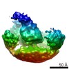 3634C  3635C  5ne4C  5nedC  5nemC  5nerC  5netC  5neuC M: map data used to model this data C: citing same article ( |
|---|---|
| Similar structure data |


-Links
 PDBj
PDBj

- Assembly
| Deposited unit | 
|
|---|---|
| 1 | x 60
|
| 2 |
|
| 3 | x 5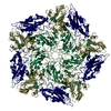
|
| 4 | x 6
|
| 5 | 
|
| Symmetry | Point symmetry: (Schoenflies symbol: I (icosahedral)) |
-Components
| #1: Protein | Mass: 23222.348 Da / Num. of mol.: 1 Source method: isolated from a genetically manipulated source Source: (gene. exp.) Foot-and-mouth disease virus / Cell line (production host): BHK-21 / Production host:  Cricetinae gen. sp. (mammal) / References: UniProt: Q6PMW3 Cricetinae gen. sp. (mammal) / References: UniProt: Q6PMW3 |
|---|---|
| #2: Protein | Mass: 24417.510 Da / Num. of mol.: 1 / Mutation: S93Y Source method: isolated from a genetically manipulated source Source: (gene. exp.) Foot-and-mouth disease virus / Cell line (production host): BHK-21 / Production host: Cricetinae gen. sp. (mammal) / References: UniProt: Q6PMW3 |
| #3: Protein | Mass: 23933.879 Da / Num. of mol.: 1 Source method: isolated from a genetically manipulated source Source: (gene. exp.) Foot-and-mouth disease virus / Cell line (production host): BHK-21 / Production host: Cricetinae gen. sp. (mammal) / References: UniProt: Q80B23, UniProt: Q6PMW3*PLUS |
| #4: Protein | Mass: 8766.075 Da / Num. of mol.: 1 Source method: isolated from a genetically manipulated source Source: (gene. exp.) Foot-and-mouth disease virus / Cell line (production host): BHK-21 / Production host: Cricetinae gen. sp. (mammal) / References: UniProt: E1ACS1, UniProt: D1H101*PLUS |
-Experimental details
-Experiment
| Experiment | Method: ELECTRON MICROSCOPY |
|---|---|
| EM experiment | Aggregation state: PARTICLE / 3D reconstruction method: single particle reconstruction |
- Sample preparation
| Component | Name: Foot-and-mouth disease virus / Type: VIRUS / Entity ID: all / Source: RECOMBINANT |
|---|---|
| Molecular weight | Value: 9 MDa / Experimental value: NO |
| Source (natural) | Organism: Foot-and-mouth disease virus / Strain: O1 Manisa |
| Source (recombinant) | Organism: Cricetinae gen. sp. (mammal) / Cell: BHK-21 |
| Details of virus | Empty: NO / Enveloped: NO / Isolate: SEROTYPE / Type: VIRION |
| Natural host | Organism: Bos taurus |
| Virus shell | Name: Foot and Mouth Disease virusFoot-and-mouth disease virus Diameter: 300 nm / Triangulation number (T number): 3 |
| Buffer solution | pH: 8 |
| Buffer component | Conc.: 50 mM / Name: HEPES |
| Specimen | Conc.: 0.5 mg/ml / Embedding applied: NO / Shadowing applied: NO / Staining applied: NO / Vitrification applied: YES |
| Specimen support | Grid material: COPPER / Grid mesh size: 300 divisions/in. / Grid type: C-flat-2/1 |
| Vitrification | Instrument: FEI VITROBOT MARK IV / Cryogen name: ETHANE / Humidity: 90 % / Chamber temperature: 294 K |
- Electron microscopy imaging
| Experimental equipment |  Model: Tecnai Polara / Image courtesy: FEI Company |
|---|---|
| Microscopy | Model: FEI POLARA 300 |
| Electron gun | Electron source: FIELD EMISSION GUN / Accelerating voltage: 300 kV / Illumination mode: FLOOD BEAM |
| Electron lens | Mode: BRIGHT FIELDBright-field microscopy / Nominal magnification: 160000 X / Calibrated magnification: 37037 X / Nominal defocus max: 4000 nm / Nominal defocus min: 1000 nm / Cs: 2 mm / C2 aperture diameter: 50 µm / Alignment procedure: BASIC |
| Specimen holder | Cryogen: NITROGEN Specimen holder model: GATAN 910 MULTI-SPECIMEN SINGLE TILT CRYO TRANSFER HOLDER Temperature (max): 70 K / Temperature (min): 70 K |
| Image recording | Average exposure time: 5 sec. / Electron dose: 18 e/Å2 / Detector mode: SUPER-RESOLUTION / Film or detector model: GATAN K2 SUMMIT (4k x 4k) / Num. of grids imaged: 1 / Num. of real images: 360 |
| EM imaging optics | Energyfilter name: GIF / Energyfilter upper: 20 eV / Energyfilter lower: 0 eV |
| Image scans | Width: 3838 / Height: 3710 / Movie frames/image: 25 / Used frames/image: 2-20 |
- Processing
| Software | Name: PHENIX / Version: dev_2645: / Classification: refinement | ||||||||||||||||||||||||||||||||||||
|---|---|---|---|---|---|---|---|---|---|---|---|---|---|---|---|---|---|---|---|---|---|---|---|---|---|---|---|---|---|---|---|---|---|---|---|---|---|
| EM software |
| ||||||||||||||||||||||||||||||||||||
| CTF correction | Type: PHASE FLIPPING AND AMPLITUDE CORRECTION | ||||||||||||||||||||||||||||||||||||
| Particle selection | Num. of particles selected: 13483 | ||||||||||||||||||||||||||||||||||||
| 3D reconstruction | Resolution: 3.1 Å / Resolution method: FSC 0.143 CUT-OFF / Num. of particles: 13483 / Algorithm: BACK PROJECTION / Symmetry type: POINT | ||||||||||||||||||||||||||||||||||||
| Atomic model building | B value: 120 / Protocol: RIGID BODY FIT / Space: REAL / Target criteria: Cross-correlation coefficient | ||||||||||||||||||||||||||||||||||||
| Atomic model building | PDB-ID: 5AC9 | ||||||||||||||||||||||||||||||||||||
| Refine LS restraints |
|