 Movie
Movie Controller


+ Open data

- Basic information
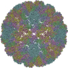
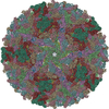
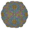
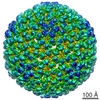
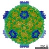
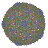
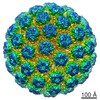
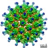
| Entry | Database: PDB / ID: 2xyy | ||||||
|---|---|---|---|---|---|---|---|
| Title | De Novo model of Bacteriophage P22 procapsid coat protein | ||||||
 Components Components | COAT PROTEIN | ||||||
 Keywords Keywords | VIRUS / ICOSAHEDRAL RECONSTRUCTION | ||||||
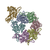
| Function / homology | Major capsid protein Gp5 / P22 coat protein - gene protein 5 / viral procapsid / viral procapsid maturation / T=7 icosahedral viral capsid / viral capsid / identical protein binding / Major capsid protein / Major capsid protein Function and homology information Function and homology information | ||||||
| Biological species |  ENTEROBACTERIA PHAGE P22 (virus) ENTEROBACTERIA PHAGE P22 (virus) | ||||||
| Method | ELECTRON MICROSCOPY / single particle reconstruction / cryo EM / Resolution: 3.8 Å | ||||||
| Model type details | CA ATOMS ONLY, CHAIN A, B, C, D, E, F, G | ||||||
 Authors Authors | Chen, D.-H. / Baker, M.L. / Hryc, C.F. / DiMaio, F. / Jakana, J. / Wu, W. / Dougherty, M. / Haase-Pettingell, C. / Schmid, M.F. / Jiang, W. ...Chen, D.-H. / Baker, M.L. / Hryc, C.F. / DiMaio, F. / Jakana, J. / Wu, W. / Dougherty, M. / Haase-Pettingell, C. / Schmid, M.F. / Jiang, W. / Baker, D. / King, J.A. / Chiu, W. | ||||||
 Citation Citation | Journal: Proc Natl Acad Sci U S A / Year: 2011 Title: Structural basis for scaffolding-mediated assembly and maturation of a dsDNA virus. Authors: Dong-Hua Chen / Matthew L Baker / Corey F Hryc / Frank DiMaio / Joanita Jakana / Weimin Wu / Matthew Dougherty / Cameron Haase-Pettingell / Michael F Schmid / Wen Jiang / David Baker / ...Authors: Dong-Hua Chen / Matthew L Baker / Corey F Hryc / Frank DiMaio / Joanita Jakana / Weimin Wu / Matthew Dougherty / Cameron Haase-Pettingell / Michael F Schmid / Wen Jiang / David Baker / Jonathan A King / Wah Chiu /  Abstract: Formation of many dsDNA viruses begins with the assembly of a procapsid, containing scaffolding proteins and a multisubunit portal but lacking DNA, which matures into an infectious virion. This ...Formation of many dsDNA viruses begins with the assembly of a procapsid, containing scaffolding proteins and a multisubunit portal but lacking DNA, which matures into an infectious virion. This process, conserved among dsDNA viruses such as herpes viruses and bacteriophages, is key to forming infectious virions. Bacteriophage P22 has served as a model system for this study in the past several decades. However, how capsid assembly is initiated, where and how scaffolding proteins bind to coat proteins in the procapsid, and the conformational changes upon capsid maturation still remain elusive. Here, we report Cα backbone models for the P22 procapsid and infectious virion derived from electron cryomicroscopy density maps determined at 3.8- and 4.0-Å resolution, respectively, and the first procapsid structure at subnanometer resolution without imposing symmetry. The procapsid structures show the scaffolding protein interacting electrostatically with the N terminus (N arm) of the coat protein through its C-terminal helix-loop-helix motif, as well as unexpected interactions between 10 scaffolding proteins and the 12-fold portal located at a unique vertex. These suggest a critical role for the scaffolding proteins both in initiating the capsid assembly at the portal vertex and propagating its growth on a T = 7 icosahedral lattice. Comparison of the procapsid and the virion backbone models reveals coordinated and complex conformational changes. These structural observations allow us to propose a more detailed molecular mechanism for the scaffolding-mediated capsid assembly initiation including portal incorporation, release of scaffolding proteins upon DNA packaging, and maturation into infectious virions. | ||||||
| History |
|
- Structure visualization
| Movie |
Movie viewer |
|---|---|
| Structure viewer | Molecule: MolmilJmol/JSmol |
- Downloads & links
-Download
| PDBx/mmCIF format | 2xyy.cif.gz | 90.5 KB | Display | PDBx/mmCIF format |
|---|---|---|---|---|
| PDB format | pdb2xyy.ent.gz | 57.4 KB | Display | PDB format |
| PDBx/mmJSON format | 2xyy.json.gz | Tree view | PDBx/mmJSON format | |
| Others |  Other downloads Other downloads |
-Validation report
| Summary document | 2xyy_validation.pdf.gz | 825.9 KB | Display | wwPDB validaton report |
|---|---|---|---|---|
| Full document | 2xyy_full_validation.pdf.gz | 825.4 KB | Display | |
| Data in XML | 2xyy_validation.xml.gz | 41.1 KB | Display | |
| Data in CIF | 2xyy_validation.cif.gz | 60.6 KB | Display | |
| Arichive directory | https://data.pdbj.org/pub/pdb/validation_reports/xy/2xyyftp://data.pdbj.org/pub/pdb/validation_reports/xy/2xyy | HTTPS FTP |
-Related structure data
| Related structure data | 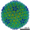 1824MC 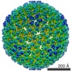 1825C 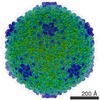 1826C 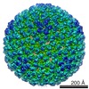 1827C 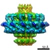 1828C 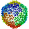 2xyzC C: citing same article ( M: map data used to model this data |
|---|---|
| Similar structure data |

-Links
 PDBj
PDBj
- Assembly
| Deposited unit | 
|
|---|---|
| 1 | x 60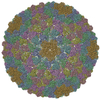
|
| 2 |
|
| 3 | x 5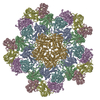
|
| 4 | x 6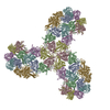
|
| 5 | 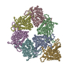
|
| Symmetry | Point symmetry: (Schoenflies symbol: I (icosahedral)) |
-Components
| #1: Protein | Mass: 46795.613 Da / Num. of mol.: 7 Source method: isolated from a genetically manipulated source Source: (gene. exp.) ENTEROBACTERIA PHAGE P22 (virus)Production host:  SALMONELLA ENTERICA SUBSP. ENTERICA SEROVAR TYPHIMURIUM (bacteria) SALMONELLA ENTERICA SUBSP. ENTERICA SEROVAR TYPHIMURIUM (bacteria)Strain (production host): DB7136 / Variant (production host): 13AMH101 / References: UniProt: A8CGC7, UniProt: P26747*PLUS |
|---|
-Experimental details
-Experiment
| Experiment | Method: ELECTRON MICROSCOPY |
|---|---|
| EM experiment | Aggregation state: PARTICLE / 3D reconstruction method: single particle reconstruction |
- Sample preparation
| Component | Name: BACTERIOPHAGE P22 PROCAPSID / Type: VIRUS / Details: BAD MICROGRAPHS WERE EXCLUDED VISUALLY |
|---|---|
| Buffer solution | Name: 50 MM TRIS PH 7.6, 25 MM NACL, 2MM EDTA / pH: 7.6 / Details: 50 MM TRIS PH 7.6, 25 MM NACL, 2MM EDTA |
| Specimen | Conc.: 1 mg/ml / Embedding applied: NO / Shadowing applied: NO / Staining applied: NO / Vitrification applied: YES |
| Specimen support | Details: HOLEY CARBON |
| Vitrification | Instrument: FEI VITROBOT MARK I / Cryogen name: ETHANE Details: VITRIFICATION 1 - CRYOGEN- ETHANE, HUMIDITY- 95, TEMPERATURE- 4.2, INSTRUMENT- VITROBOT, METHOD- BLOT FOR 2 SECONDS BEFORE PLUNGING, |
- Electron microscopy imaging
| Microscopy | Model: JEOL KYOTO-3000SFF / Date: Mar 7, 2008 |
|---|---|
| Electron gun | Electron source:  FIELD EMISSION GUN / Accelerating voltage: 300 kV / Illumination mode: FLOOD BEAM FIELD EMISSION GUN / Accelerating voltage: 300 kV / Illumination mode: FLOOD BEAM |
| Electron lens | Mode: BRIGHT FIELD / Nominal magnification: 60000 X / Calibrated magnification: 60000 X / Nominal defocus max: 2800 nm / Nominal defocus min: 500 nm / Cs: 1.6 mm |
| Specimen holder | Temperature: 4.2 K |
| Image recording | Electron dose: 36 e/Å2 / Film or detector model: KODAK SO-163 FILM |
| Image scans | Num. digital images: 921 |
- Processing
| EM software | Name: EMAN / Category: 3D reconstruction | ||||||||||||
|---|---|---|---|---|---|---|---|---|---|---|---|---|---|
| CTF correction | Details: EACH PARTICLE | ||||||||||||
| Symmetry | Point symmetry: I (icosahedral) | ||||||||||||
| 3D reconstruction | Method: FOURIER METHODS / Resolution: 3.8 Å / Num. of particles: 23400 / Nominal pixel size: 1.06 Å / Actual pixel size: 1.06 Å Details: MAKE3D IN EMAN.N-TERMINAL 9 RESIDUES AND C-TERMINAL 5 RESIDUES WERE NOT MODELED.SUBMISSION BASED ON EXPERIMENTAL DATA FROM EMDB EMD-1824. Symmetry type: POINT | ||||||||||||
| Atomic model building | Protocol: OTHER / Space: REAL / Target criteria: FSC / Details: REFINEMENT PROTOCOL--EM | ||||||||||||
| Refinement | Highest resolution: 3.8 Å | ||||||||||||
| Refinement step | Cycle: LAST / Highest resolution: 3.8 Å
|
