 Movie
Movie Controller

[English] 日本語
 Yorodumi
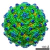
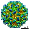
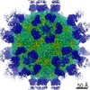
- EMDB-2027: Coxsackievirus A7 (CAV7) empty capsid reconstruction at 6.09 angs... -
+ Open data

- Basic information
| Entry | Database: EMDB / ID: EMD-2027 | |||||||||
|---|---|---|---|---|---|---|---|---|---|---|
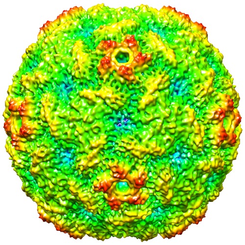
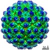
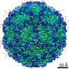
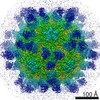
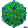
| Title | Coxsackievirus A7 (CAV7) empty capsid reconstruction at 6.09 angstrom resolution. | |||||||||
 Map data Map data | This is a three-dimensional model of coxsackievirus A7 (CAV7) at 6.09 angstrom resolution. | |||||||||
 Sample Sample |
| |||||||||
 Keywords Keywords | coxsackievirus / CAV7 / picornavirus / enterovirus / HEV-A | |||||||||
| Function / homology |  Function and homology information Function and homology informationviral capsid / symbiont entry into host cell / virion attachment to host cell / structural molecule activity Similarity search - Function | |||||||||
| Biological species |  Human coxsackievirus A7 Human coxsackievirus A7 | |||||||||
| Method | single particle reconstruction / cryo EM / Resolution: 6.09 Å | |||||||||
 Authors Authors | Seitsonen JJT / Shakeel S / Susi P / Pandurangan AP / Sinkovits RS / Hyvonen H / Laurinmaki P / Yla-Pelto J / Topf M / Hyypia T / Butcher SJ | |||||||||
 Citation Citation | Journal: J Virol / Year: 2012 Title: Structural analysis of coxsackievirus A7 reveals conformational changes associated with uncoating. Authors: Jani J T Seitsonen / Shabih Shakeel / Petri Susi / Arun P Pandurangan / Robert S Sinkovits / Heini Hyvönen / Pasi Laurinmäki / Jani Ylä-Pelto / Maya Topf / Timo Hyypiä / Sarah J Butcher /  Abstract: Coxsackievirus A7 (CAV7) is a rarely detected and poorly characterized serotype of the Enterovirus species Human enterovirus A (HEV-A) within the Picornaviridae family. The CAV7-USSR strain has ...Coxsackievirus A7 (CAV7) is a rarely detected and poorly characterized serotype of the Enterovirus species Human enterovirus A (HEV-A) within the Picornaviridae family. The CAV7-USSR strain has caused polio-like epidemics and was originally thought to represent the fourth poliovirus type, but later evidence linked this strain to the CAV7-Parker prototype. Another isolate, CAV7-275/58, was also serologically similar to Parker but was noninfectious in a mouse model. Sequencing of the genomic region encoding the capsid proteins of the USSR and 275/58 strains and subsequent comparison with the corresponding amino acid sequences of the Parker strain revealed that the Parker and USSR strains are nearly identical, while the 275/58 strain is more distant. Using electron cryomicroscopy and three-dimensional image reconstruction, the structures of the CAV7-USSR virion and empty capsid were resolved to 8.2-Å and 6.1-Å resolutions, respectively. This is one of the first detailed structural analyses of the HEV-A species. Using homology modeling, reconstruction segmentation, and flexible fitting, we constructed a pseudoatomic T = 1 (pseudo T = 3) model incorporating the three major capsid proteins (VP1 to VP3), addressed the conformational changes of the capsid and its constituent viral proteins occurring during RNA release, and mapped the capsid proteins' variable regions to the structure. During uncoating, VP4 and RNA are released analogously to poliovirus 1, the interfaces of VP2 and VP3 are rearranged, and VP1 rotates. Variable regions in the capsid proteins were predicted to map mainly to the surface of VP1 and are thus likely to affect the tropism and pathogenicity of CAV7. | |||||||||
| History |
|
- Structure visualization
| Movie |
Movie viewer |
|---|---|
| Structure viewer | EM map: SurfViewMolmilJmol/JSmol |
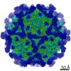
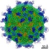
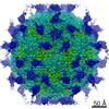
| Supplemental images |
- Downloads & links
-EMDB archive
| Map data | emd_2027.map.gz | 53.4 MB | EMDB map data format | |
|---|---|---|---|---|
| Header (meta data) | emd-2027-v30.xmlemd-2027.xml | 9.3 KB 9.3 KB | Display Display | EMDB header |
| Images | EM-2027.tif | 526.8 KB | ||
| Archive directory |  http://ftp.pdbj.org/pub/emdb/structures/EMD-2027ftp://ftp.pdbj.org/pub/emdb/structures/EMD-2027 http://ftp.pdbj.org/pub/emdb/structures/EMD-2027ftp://ftp.pdbj.org/pub/emdb/structures/EMD-2027 | HTTPS FTP |
-Validation report
| Summary document | emd_2027_validation.pdf.gz | 293.9 KB | Display | EMDB validaton report |
|---|---|---|---|---|
| Full document | emd_2027_full_validation.pdf.gz | 293.1 KB | Display | |
| Data in XML | emd_2027_validation.xml.gz | 6.5 KB | Display | |
| Arichive directory | https://ftp.pdbj.org/pub/emdb/validation_reports/EMD-2027ftp://ftp.pdbj.org/pub/emdb/validation_reports/EMD-2027 | HTTPS FTP |
-Related structure data
| Related structure data |  4biqM  2028C M: atomic model generated by this map C: citing same article ( |
|---|---|
| Similar structure data |
-Links
| EMDB pages | EMDB (EBI/PDBe) / EMDataResource |
|---|---|
| Related items in Molecule of the Month |

-Map
| File | Download / File: emd_2027.map.gz / Format: CCP4 / Size: 120.1 MB / Type: IMAGE STORED AS SIGNED INTEGER (2 BYTES) | ||||||||||||||||||||||||||||||||||||||||||||||||||||||||||||||||||||
|---|---|---|---|---|---|---|---|---|---|---|---|---|---|---|---|---|---|---|---|---|---|---|---|---|---|---|---|---|---|---|---|---|---|---|---|---|---|---|---|---|---|---|---|---|---|---|---|---|---|---|---|---|---|---|---|---|---|---|---|---|---|---|---|---|---|---|---|---|---|
| Annotation | This is a three-dimensional model of coxsackievirus A7 (CAV7) at 6.09 angstrom resolution. | ||||||||||||||||||||||||||||||||||||||||||||||||||||||||||||||||||||
| Voxel size | X=Y=Z: 1.13 Å | ||||||||||||||||||||||||||||||||||||||||||||||||||||||||||||||||||||
| Density |
| ||||||||||||||||||||||||||||||||||||||||||||||||||||||||||||||||||||
| Symmetry | Space group: 1 | ||||||||||||||||||||||||||||||||||||||||||||||||||||||||||||||||||||
| Details | EMDB XML:
CCP4 map header:
| ||||||||||||||||||||||||||||||||||||||||||||||||||||||||||||||||||||
-Supplemental data
- Sample components
-Entire : Coxsackievirus A7 USSR-strain empty particles in buffer
| Entire | Name: Coxsackievirus A7 USSR-strain empty particles in buffer |
|---|---|
| Components |
|
-Supramolecule #1000: Coxsackievirus A7 USSR-strain empty particles in buffer
| Supramolecule | Name: Coxsackievirus A7 USSR-strain empty particles in buffer type: sample / ID: 1000 / Oligomeric state: empty particle (no RNA) / Number unique components: 1 |
|---|---|
| Molecular weight | Theoretical: 5.2 MDa |
-Supramolecule #1: Human coxsackievirus A7
| Supramolecule | Name: Human coxsackievirus A7 / type: virus / ID: 1 / Name.synonym: CAV7 / NCBI-ID: 42787 / Sci species name: Human coxsackievirus A7 / Virus type: VIRION / Virus isolate: STRAIN / Virus enveloped: No / Virus empty: Yes / Syn species name: CAV7 |
|---|---|
| Host (natural) | Organism:  Homo sapiens (human) / synonym: VERTEBRATES Homo sapiens (human) / synonym: VERTEBRATES |
| Molecular weight | Theoretical: 5.2 MDa |
| Virus shell | Shell ID: 1 / Name: VP1VP2VP3 / Diameter: 300 Å / T number (triangulation number): 1 |
-Experimental details
-Structure determination
| Method | cryo EM |
|---|---|
 Processing Processing | single particle reconstruction |
| Aggregation state | particle |
-Sample preparation
| Concentration | 3 mg/mL |
|---|---|
| Buffer | pH: 7.4 / Details: 25 mM phosphate buffer, 0.5 mM MgCl2 |
| Vitrification | Cryogen name: ETHANE / Instrument: HOMEMADE PLUNGER / Details: Vitrification instrument: Custom built / Method: Manual |
- Electron microscopy
| Microscope | FEI TECNAI F20 |
|---|---|
| Image recording | Category: FILM / Film or detector model: KODAK SO-163 FILM / Digitization - Scanner: OTHER / Digitization - Sampling interval: 7 µm / Number real images: 264 / Average electron dose: 17 e/Å2 / Details: Scanned with Zeiss Photoscan TD |
| Electron beam | Acceleration voltage: 200 kV / Electron source:  FIELD EMISSION GUN FIELD EMISSION GUN |
| Electron optics | Illumination mode: FLOOD BEAM / Imaging mode: BRIGHT FIELD / Cs: 2.0 mm / Nominal defocus max: 4.81 µm / Nominal defocus min: 0.62 µm / Nominal magnification: 62000 |
| Sample stage | Specimen holder: Gatan 626 / Specimen holder model: GATAN LIQUID NITROGEN |
| Experimental equipment |  Model: Tecnai F20 / Image courtesy: FEI Company |
-Image processing
| CTF correction | Details: Each micrograph |
|---|---|
| Final reconstruction | Applied symmetry - Point group: I (icosahedral) / Algorithm: OTHER / Resolution.type: BY AUTHOR / Resolution: 6.09 Å / Resolution method: FSC 0.5 CUT-OFF / Software - Name: AUTO3DEM / Number images used: 7849 |
