Movie
Movie Controller

[English] 日本語
 Yorodumi
- EMDB-5369: Structural transitions in RCNMV revealing potential mechanism of ... -
+ Open data

- Basic information
| Entry | Database: EMDB / ID: EMD-5369 | |||||||||
|---|---|---|---|---|---|---|---|---|---|---|
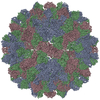
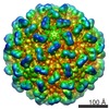
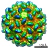
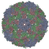
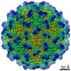
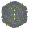
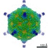
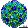
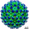
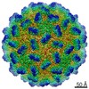
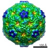
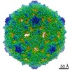
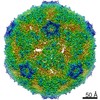
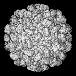
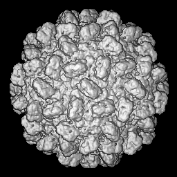
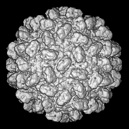
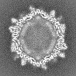
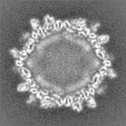
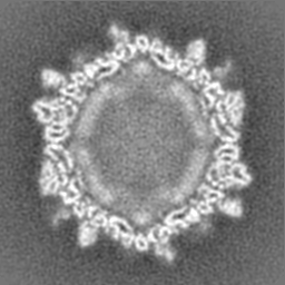
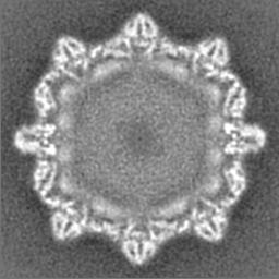
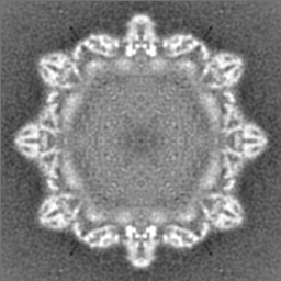
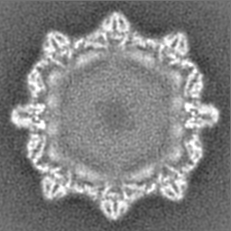
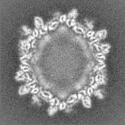
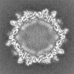
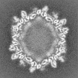
| Title | Structural transitions in RCNMV revealing potential mechanism of RNA release (native map) | |||||||||
 Map data Map data | this is EM map of native RCNMV | |||||||||
 Sample Sample |
| |||||||||
 Keywords Keywords | cryo-electron microscopy / virus / red clover necrotic mosaic virus | |||||||||
| Biological species |  Red clover necrotic mosaic virus Red clover necrotic mosaic virus | |||||||||
| Method | single particle reconstruction / cryo EM / Resolution: 8.5 Å | |||||||||
 Authors Authors | Sherman MB / Guenther RH / Tama F / Sit TL / Brooks CL / Mikhailov AM / Orlova EV / Baker TS / Lommel SA | |||||||||
 Citation Citation | Journal: J Virol / Year: 2006 Title: Removal of divalent cations induces structural transitions in red clover necrotic mosaic virus, revealing a potential mechanism for RNA release. Authors: Michael B Sherman / Richard H Guenther / Florence Tama / Tim L Sit / Charles L Brooks / Albert M Mikhailov / Elena V Orlova / Timothy S Baker / Steven A Lommel /  Abstract: The structure of Red clover necrotic mosaic virus (RCNMV), an icosahedral plant virus, was resolved to 8.5 A by cryoelectron microscopy. The virion capsid has prominent surface protrusions and ...The structure of Red clover necrotic mosaic virus (RCNMV), an icosahedral plant virus, was resolved to 8.5 A by cryoelectron microscopy. The virion capsid has prominent surface protrusions and subunits with a clearly defined shell and protruding domains. The structures of both the individual capsid protein (CP) subunits and the entire virion capsid are consistent with other species in the Tombusviridae family. Within the RCNMV capsid, there is a clearly defined inner cage formed by complexes of genomic RNA and the amino termini of CP subunits. An RCNMV virion has approximately 390 +/- 30 Ca2+ ions bound to the capsid and 420 +/- 25 Mg2+ ions thought to be in the interior of the capsid. Depletion of both Ca2+ and Mg2+ ions from RCNMV leads to significant structural changes, including (i) formation of 11- to 13-A-diameter channels that extend through the capsid and (ii) significant reorganization within the interior of the capsid. Genomic RNA within native capsids containing both Ca2+ and Mg2+ ions is extremely resistant to nucleases, but depletion of both of these cations results in nuclease sensitivity, as measured by a significant reduction in RCNMV infectivity. These results indicate that divalent cations play a central role in capsid dynamics and suggest a mechanism for the release of viral RNA in low-divalent-cation environments such as those found within the cytoplasm of a cell. | |||||||||
| History |
|
- Structure visualization
| Movie |
 Movie viewer Movie viewer |
|---|---|
| Structure viewer | EM map: SurfViewMolmilJmol/JSmol |
| Supplemental images |

- Downloads & links
-EMDB archive
| Map data | emd_5369.map.gz | 30.9 MB | EMDB map data format | |
|---|---|---|---|---|
| Header (meta data) | emd-5369-v30.xmlemd-5369.xml | 10.9 KB 10.9 KB | Display Display | EMDB header |
| Images | 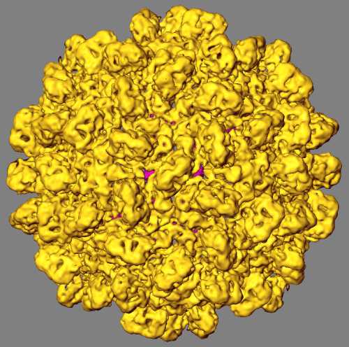 emd_5369_1.jpg emd_5369_1.jpg | 39.6 KB | ||
| Archive directory |  http://ftp.pdbj.org/pub/emdb/structures/EMD-5369ftp://ftp.pdbj.org/pub/emdb/structures/EMD-5369 http://ftp.pdbj.org/pub/emdb/structures/EMD-5369ftp://ftp.pdbj.org/pub/emdb/structures/EMD-5369 | HTTPS FTP |
-Related structure data
-Links
| EMDB pages | EMDB (EBI/PDBe) / EMDataResource |
|---|
-Map
| File | Download / File: emd_5369.map.gz / Format: CCP4 / Size: 62.5 MB / Type: IMAGE STORED AS FLOATING POINT NUMBER (4 BYTES) | ||||||||||||||||||||||||||||||||||||||||||||||||||||||||||||||||||||
|---|---|---|---|---|---|---|---|---|---|---|---|---|---|---|---|---|---|---|---|---|---|---|---|---|---|---|---|---|---|---|---|---|---|---|---|---|---|---|---|---|---|---|---|---|---|---|---|---|---|---|---|---|---|---|---|---|---|---|---|---|---|---|---|---|---|---|---|---|---|
| Annotation | this is EM map of native RCNMV | ||||||||||||||||||||||||||||||||||||||||||||||||||||||||||||||||||||
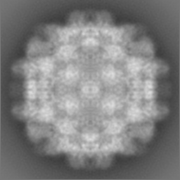
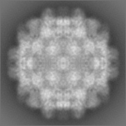
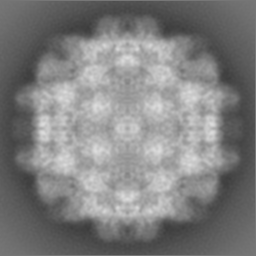
| Projections & slices | Image control
Images are generated by Spider. | ||||||||||||||||||||||||||||||||||||||||||||||||||||||||||||||||||||
| Voxel size | X=Y=Z: 1.5 Å | ||||||||||||||||||||||||||||||||||||||||||||||||||||||||||||||||||||
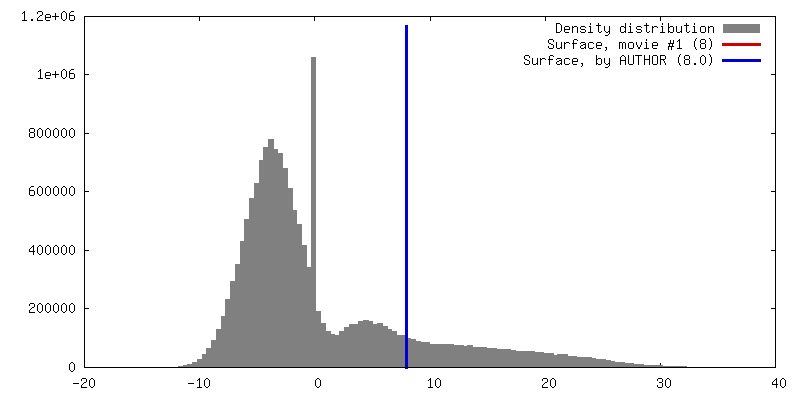
| Density |
| ||||||||||||||||||||||||||||||||||||||||||||||||||||||||||||||||||||
| Symmetry | Space group: 1 | ||||||||||||||||||||||||||||||||||||||||||||||||||||||||||||||||||||
| Details | EMDB XML:
CCP4 map header:
| ||||||||||||||||||||||||||||||||||||||||||||||||||||||||||||||||||||
 Z (Sec.)
 Y (Row.)
 X (Col.)


-Supplemental data
- Sample components
-Entire : RCNMV
| Entire | Name: RCNMV |
|---|---|
| Components |
|
-Supramolecule #1000: RCNMV
| Supramolecule | Name: RCNMV / type: sample / ID: 1000 / Details: monodisperse sample / Number unique components: 1 |
|---|---|
| Molecular weight | Theoretical: 7 MDa |
-Supramolecule #1: Red clover necrotic mosaic virus
| Supramolecule | Name: Red clover necrotic mosaic virus / type: virus / ID: 1 / Name.synonym: RCNMV / NCBI-ID: 12267 / Sci species name: Red clover necrotic mosaic virus / Database: NCBI / Virus type: VIRION / Virus isolate: SPECIES / Virus enveloped: No / Virus empty: No / Syn species name: RCNMV |
|---|---|
| Host (natural) | Organism:  Dianthus (plant) / synonym: PLANTAE(HIGHER PLANTS) Dianthus (plant) / synonym: PLANTAE(HIGHER PLANTS) |
| Molecular weight | Experimental: 7 MDa / Theoretical: 7 MDa |
| Virus shell | Shell ID: 1 / Diameter: 366 Å / T number (triangulation number): 3 |
-Experimental details
-Structure determination
| Method | cryo EM |
|---|---|
 Processing Processing | single particle reconstruction |
| Aggregation state | particle |
-Sample preparation
| Concentration | 1 mg/mL |
|---|---|
| Buffer | pH: 6.5 / Details: 10 mM Tris-HCl |
| Grid | Details: 200 mesh Quantifoil |
| Vitrification | Cryogen name: ETHANE / Chamber temperature: 90 K / Instrument: HOMEMADE PLUNGER / Details: Vitrification instrument: cryo-plunger |
- Electron microscopy
| Microscope | FEI/PHILIPS CM300FEG/T |
|---|---|
| Temperature | Min: 95 K / Max: 123 K / Average: 95 K |
| Alignment procedure | Legacy - Astigmatism: manual / Legacy - Electron beam tilt params: 0 |
| Date | Oct 15, 2003 |
| Image recording | Category: FILM / Film or detector model: KODAK SO-163 FILM / Digitization - Scanner: ZEISS SCAI / Digitization - Sampling interval: 7 µm / Number real images: 2500 / Average electron dose: 20 e/Å2 / Od range: 1.5 / Bits/pixel: 16 |
| Tilt angle min | 0 |
| Tilt angle max | 0 |
| Electron beam | Acceleration voltage: 300 kV / Electron source:  FIELD EMISSION GUN FIELD EMISSION GUN |
| Electron optics | Calibrated magnification: 49523 / Illumination mode: FLOOD BEAM / Imaging mode: BRIGHT FIELD / Cs: 2.0 mm / Nominal defocus max: 2.8 µm / Nominal defocus min: 0.8 µm / Nominal magnification: 47000 |
| Sample stage | Specimen holder: GATAN 626 / Specimen holder model: GATAN LIQUID NITROGEN |
-Image processing
| CTF correction | Details: micrograph |
|---|---|
| Final reconstruction | Algorithm: OTHER / Resolution.type: BY AUTHOR / Resolution: 8.5 Å / Resolution method: FSC 0.5 CUT-OFF / Software - Name: PFT / Number images used: 4000 |
-Atomic model buiding 1
| Initial model | PDB ID: |
|---|---|
| Software | Name: SITUS |
| Details | Protocol: NNM |
| Refinement | Space: REAL |