 Movie
Movie Controller

[English] 日本語
 Yorodumi
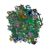
- PDB-5ojq: The modeled structure of of wild type extended type VI secretion ... -
+ Open data

- Basic information
| Entry | Database: PDB / ID: 5ojq | ||||||
|---|---|---|---|---|---|---|---|
| Title | The modeled structure of of wild type extended type VI secretion system sheath/tube complex in vibrio cholerae based on cryo-EM reconstruction of the non-contractile sheath/tube complex | ||||||
 Components Components |
| ||||||
 Keywords Keywords | STRUCTURAL PROTEIN / T6SS / extended conformation / vibrio cholerae | ||||||
| Function / homology |  Function and homology information Function and homology informationType VI secretion system TssC-like / TssC1, N-terminal / TssC1, C-terminal / EvpB/VC_A0108, tail sheath N-terminal domain / EvpB/VC_A0108, tail sheath gpW/gp25-like domain / Type VI secretion system sheath protein TssB1 / Type VI secretion system, VipA, VC_A0107 or Hcp2 / Type VI secretion system effector Hcp / Hcp1-like superfamily / Type VI secretion system effector, Hcp Similarity search - Domain/homology | ||||||
| Biological species |   Vibrio cholerae (bacteria) Vibrio cholerae (bacteria) | ||||||
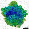
| Method | ELECTRON MICROSCOPY / helical reconstruction / cryo EM / Resolution: 3.7 Å | ||||||
 Authors Authors | Wang, J. / Brackmann, M. / Castano-Diez, D. / Kudryashev, M. / Goldie, K. / Maier, T. / Stahlberg, H. / Basler, M. | ||||||
| Funding support |  Switzerland, 1items Switzerland, 1items
| ||||||
 Citation Citation | Journal: Nat Microbiol / Year: 2017 Title: Cryo-EM structure of the extended type VI secretion system sheath-tube complex. Authors: Jing Wang / Maximilian Brackmann / Daniel Castaño-Díez / Mikhail Kudryashev / Kenneth N Goldie / Timm Maier / Henning Stahlberg / Marek Basler /  Abstract: The bacterial type VI secretion system (T6SS) uses contraction of a long sheath to quickly thrust a tube with associated effectors across membranes of eukaryotic and bacterial cells . Only limited ...The bacterial type VI secretion system (T6SS) uses contraction of a long sheath to quickly thrust a tube with associated effectors across membranes of eukaryotic and bacterial cells . Only limited structural information is available about the inherently unstable precontraction state of the T6SS. Here, we obtain a 3.7 Å resolution structure of a non-contractile sheath-tube complex using cryo-electron microscopy and show that it resembles the extended T6SS inside Vibrio cholerae cells. We build a pseudo-atomic model of the complete sheath-tube assembly, which provides a mechanistic understanding of coupling sheath contraction with pushing and rotating the inner tube for efficient target membrane penetration. Our data further show that sheath contraction exposes a buried recognition domain to specifically trigger the disassembly and recycling of the T6SS sheath by the cognate ATP-dependent unfoldase ClpV. | ||||||
| History |
|
- Structure visualization
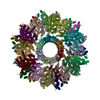
| Movie |
Movie viewer |
|---|---|
| Structure viewer | Molecule: MolmilJmol/JSmol |
- Downloads & links
-Download
| PDBx/mmCIF format | 5ojq.cif.gz | 2.3 MB | Display | PDBx/mmCIF format |
|---|---|---|---|---|
| PDB format | pdb5ojq.ent.gz | Display | PDB format | |
| PDBx/mmJSON format | 5ojq.json.gz | Tree view | PDBx/mmJSON format | |
| Others |  Other downloads Other downloads |
-Validation report
| Summary document | 5ojq_validation.pdf.gz | 1.2 MB | Display | wwPDB validaton report |
|---|---|---|---|---|
| Full document | 5ojq_full_validation.pdf.gz | 1.3 MB | Display | |
| Data in XML | 5ojq_validation.xml.gz | 253.9 KB | Display | |
| Data in CIF | 5ojq_validation.cif.gz | 442.9 KB | Display | |
| Arichive directory | https://data.pdbj.org/pub/pdb/validation_reports/oj/5ojqftp://data.pdbj.org/pub/pdb/validation_reports/oj/5ojq | HTTPS FTP |
-Related structure data
| Related structure data | 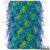 3566MC 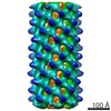 3563C 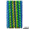 3564C 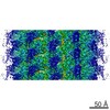 3567C 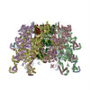 5mxnC 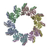 5myuC M: map data used to model this data C: citing same article ( |
|---|---|
| Similar structure data | |
| EM raw data | EMPIAR-10086 (Title: Cryo EM of Type VI Secretion System VipA-N3/VipB/Hcp complex Data size: 26.2 Data #1: Unaligned multi-frame micrographs of T6SS VipA-N3/VipB/Hcp complex [micrographs - multiframe]) |

-Links
 PDBj
PDBj- Assembly
| Deposited unit | 
|
|---|---|
| 1 |
|
| Details | details |
-Components
| #1: Protein | Mass: 18878.055 Da / Num. of mol.: 18 / Source method: isolated from a natural source / Source: (natural) Vibrio cholerae (bacteria) / References: UniProt: P72350#2: Protein | Mass: 53504.434 Da / Num. of mol.: 18 / Source method: isolated from a natural source / Source: (natural) Vibrio cholerae (bacteria) / References: UniProt: A0A085SGI6, UniProt: Q9KN57*PLUS#3: Protein | Mass: 17099.416 Da / Num. of mol.: 18 / Source method: isolated from a natural source / Source: (natural) Vibrio cholerae (bacteria) / References: UniProt: A0A023PRF3, UniProt: Q9KN58*PLUS |
|---|
-Experimental details
-Experiment
| Experiment | Method: ELECTRON MICROSCOPY |
|---|---|
| EM experiment | Aggregation state: HELICAL ARRAY / 3D reconstruction method: helical reconstruction |
- Sample preparation
| Component | Name: T6SS extended sheath/tube complex / Type: COMPLEX / Entity ID: all / Source: NATURAL |
|---|---|
| Source (natural) | Organism: Vibrio cholerae (bacteria) |
| Buffer solution | pH: 7.5 |
| Specimen | Embedding applied: NO / Shadowing applied: NO / Staining applied: NO / Vitrification applied: YES |
| Vitrification | Cryogen name: ETHANE |
- Electron microscopy imaging
| Experimental equipment |  Model: Titan Krios / Image courtesy: FEI Company |
|---|---|
| Microscopy | Model: FEI TITAN KRIOS |
| Electron gun | Electron source:  FIELD EMISSION GUN / Accelerating voltage: 300 kV / Illumination mode: FLOOD BEAM FIELD EMISSION GUN / Accelerating voltage: 300 kV / Illumination mode: FLOOD BEAM |
| Electron lens | Mode: BRIGHT FIELD |
| Image recording | Electron dose: 1 e/Å2 / Film or detector model: GATAN K2 SUMMIT (4k x 4k) |
- Processing
| CTF correction | Type: PHASE FLIPPING AND AMPLITUDE CORRECTION |
|---|---|
| Helical symmerty | Angular rotation/subunit: 23.5 ° / Axial rise/subunit: 37.8 Å / Axial symmetry: C6 |
| 3D reconstruction | Resolution: 3.7 Å / Resolution method: FSC 0.143 CUT-OFF / Num. of particles: 10000 / Symmetry type: HELICAL |
