 Movie
Movie Controller

[English] 日本語
 Yorodumi
- PDB-5urw: Structure of the extended type VI secretion system sheath in Myxo... -
+ Open data

- Basic information
| Entry | Database: PDB / ID: 5urw | ||||||
|---|---|---|---|---|---|---|---|
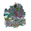
| Title | Structure of the extended type VI secretion system sheath in Myxococcus xanthus | ||||||
 Components Components |
| ||||||
 Keywords Keywords | PROTEIN TRANSPORT / T6SS / type IV secretion / protein machine / secretion system / sheath / contractile | ||||||
| Function / homology |  Function and homology information Function and homology information: / Type VI secretion system TssC-like / TssC1, N-terminal / TssC1, C-terminal / EvpB/VC_A0108, tail sheath N-terminal domain / EvpB/VC_A0108, tail sheath gpW/gp25-like domain / Type VI secretion system sheath protein TssB1 / Type VI secretion system, VipA, VC_A0107 or Hcp2 / Type VI secretion system effector Hcp / Hcp1-like superfamily / Type VI secretion system effector, Hcp Similarity search - Domain/homology | ||||||
| Biological species |  Myxococcus xanthus (bacteria) Myxococcus xanthus (bacteria) | ||||||
| Method | ELECTRON MICROSCOPY / subtomogram averaging / cryo EM / Resolution: 24 Å | ||||||
 Authors Authors | Chang, Y.-W. / Rettberg, L.A. / Jensen, G.J. | ||||||
 Citation Citation | Journal: EMBO Rep / Year: 2017 Title: structures of an intact type VI secretion system revealed by electron cryotomography. Authors: Yi-Wei Chang / Lee A Rettberg / Davi R Ortega / Grant J Jensen /  Abstract: The type VI secretion system (T6SS) is a versatile molecular weapon used by many bacteria against eukaryotic hosts or prokaryotic competitors. It consists of a cytoplasmic bacteriophage tail-like ...The type VI secretion system (T6SS) is a versatile molecular weapon used by many bacteria against eukaryotic hosts or prokaryotic competitors. It consists of a cytoplasmic bacteriophage tail-like structure anchored in the bacterial cell envelope via a cytoplasmic baseplate and a periplasmic membrane complex. Rapid contraction of the sheath in the bacteriophage tail-like structure propels an inner tube/spike complex through the target cell envelope to deliver effectors. While structures of purified contracted sheath and purified membrane complex have been solved, because sheaths contract upon cell lysis and purification, no structure is available for the extended sheath. Structural information about the baseplate is also lacking. Here, we use electron cryotomography to directly visualize intact T6SS structures inside cells. Using sub-tomogram averaging, we resolve the structure of the extended sheath and membrane-associated components including the baseplate. Moreover, we identify novel extracellular bacteriophage tail fiber-like antennae. These results provide new structural insights into how the extended sheath prevents premature disassembly and how this sophisticated machine may recognize targets. | ||||||
| History |
|
- Structure visualization
| Movie |
Movie viewer |
|---|---|
| Structure viewer | Molecule: MolmilJmol/JSmol |
- Downloads & links
-Download
| PDBx/mmCIF format | 5urw.cif.gz | 1.5 MB | Display | PDBx/mmCIF format |
|---|---|---|---|---|
| PDB format | pdb5urw.ent.gz | Display | PDB format | |
| PDBx/mmJSON format | 5urw.json.gz | Tree view | PDBx/mmJSON format | |
| Others |  Other downloads Other downloads |
-Validation report
| Arichive directory | https://data.pdbj.org/pub/pdb/validation_reports/ur/5urwftp://data.pdbj.org/pub/pdb/validation_reports/ur/5urw | HTTPS FTP |
|---|
-Related structure data
| Related structure data | 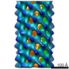 8601MC 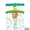 8600C 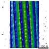 8602C 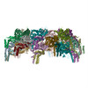 5urxC M: map data used to model this data C: citing same article ( |
|---|---|
| Similar structure data |

-Links
 PDBj
PDBj- Assembly
| Deposited unit | 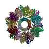
|
|---|---|
| 1 |
|
-Components
| #1: Protein | Mass: 18016.617 Da / Num. of mol.: 18 / Source method: isolated from a natural source Source: (natural) Myxococcus xanthus (strain DK 1622) (bacteria)Strain: DK 1622 / References: UniProt: Q1D305 #2: Protein | Mass: 56188.469 Da / Num. of mol.: 18 / Source method: isolated from a natural source / Source: (natural) Myxococcus xanthus (bacteria) / Strain: DK 1622 / References: UniProt: Q1D304#3: Protein | Mass: 18065.271 Da / Num. of mol.: 18 / Source method: isolated from a natural source / Source: (natural) Myxococcus xanthus (bacteria) / Strain: DK 1622 / References: UniProt: Q1D303 |
|---|
-Experimental details
-Experiment
| Experiment | Method: ELECTRON MICROSCOPY |
|---|---|
| EM experiment | Aggregation state: CELL / 3D reconstruction method: subtomogram averaging |
- Sample preparation
| Component | Name: Type VI secretion system / Type: COMPLEX / Entity ID: all / Source: NATURAL |
|---|---|
| Source (natural) | Organism: Myxococcus xanthus DK 1622 (bacteria) |
| Buffer solution | pH: 7 |
| Specimen | Embedding applied: NO / Shadowing applied: NO / Staining applied: NO / Vitrification applied: YES |
| Vitrification | Cryogen name: ETHANE-PROPANE |
- Electron microscopy imaging
| Experimental equipment |  Model: Tecnai Polara / Image courtesy: FEI Company |
|---|---|
| Microscopy | Model: FEI POLARA 300 |
| Electron gun | Electron source:  FIELD EMISSION GUN / Accelerating voltage: 300 kV / Illumination mode: FLOOD BEAM FIELD EMISSION GUN / Accelerating voltage: 300 kV / Illumination mode: FLOOD BEAM |
| Electron lens | Mode: BRIGHT FIELD |
| Image recording | Electron dose: 1.5 e/Å2 / Film or detector model: GATAN K2 SUMMIT (4k x 4k) |
- Processing
| EM software |
| ||||||||||||||||||
|---|---|---|---|---|---|---|---|---|---|---|---|---|---|---|---|---|---|---|---|
| CTF correction | Type: NONE | ||||||||||||||||||
| 3D reconstruction | Resolution: 24 Å / Resolution method: OTHER / Num. of particles: 687 / Symmetry type: POINT | ||||||||||||||||||
| EM volume selection | Num. of tomograms: 29 / Num. of volumes extracted: 687 |
