 Movie
Movie Controller


+ Open data

- Basic information
| Entry | Database: PDB / ID: 4urd | ||||||
|---|---|---|---|---|---|---|---|
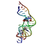
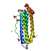
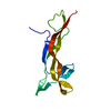
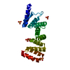
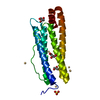
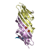
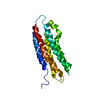
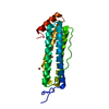
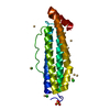
| Title | Cryo-EM map of Trigger Factor bound to a translating ribosome | ||||||
 Components Components | TRIGGER FACTOR | ||||||
 Keywords Keywords | ISOMERASE / TRANSLATION / CO-TRANSLATIONAL PROTEIN FOLDING | ||||||
| Function / homology |  Function and homology information Function and homology information'de novo' cotranslational protein folding / stress response to copper ion / protein unfolding / chaperone-mediated protein folding / protein folding chaperone / peptidylprolyl isomerase / peptidyl-prolyl cis-trans isomerase activity / protein transport / ribosome binding / response to heat ...'de novo' cotranslational protein folding / stress response to copper ion / protein unfolding / chaperone-mediated protein folding / protein folding chaperone / peptidylprolyl isomerase / peptidyl-prolyl cis-trans isomerase activity / protein transport / ribosome binding / response to heat / cell division / identical protein binding / membrane / cytosol Similarity search - Function | ||||||
| Biological species |  | ||||||
| Method | ELECTRON MICROSCOPY / single particle reconstruction / cryo EM / Resolution: 7.7 Å | ||||||
 Authors Authors | Deeng, J. / Chan, K.Y. / van der Sluis, E. / Bischoff, L. / Berninghausen, O. / Han, W. / Gumbart, J. / Schulten, K. / Beatrix, B. / Beckmann, R. | ||||||
 Citation Citation | Journal: J Mol Biol / Year: 2016 Title: Dynamic Behavior of Trigger Factor on the Ribosome. Authors: J Deeng / K Y Chan / E O van der Sluis / O Berninghausen / W Han / J Gumbart / K Schulten / B Beatrix / R Beckmann /   Abstract: Trigger factor (TF) is the only ribosome-associated chaperone in bacteria. It interacts with hydrophobic segments in nascent chain (NCs) as they emerge from the ribosome. TF binds via its N-terminal ...Trigger factor (TF) is the only ribosome-associated chaperone in bacteria. It interacts with hydrophobic segments in nascent chain (NCs) as they emerge from the ribosome. TF binds via its N-terminal ribosome-binding domain (RBD) mainly to ribosomal protein uL23 at the tunnel exit on the large ribosomal subunit. Whereas earlier structural data suggested that TF binds as a rigid molecule to the ribosome, recent comparisons of structural data on substrate-bound, ribosome-bound, and TF in solution from different species suggest that this chaperone is a rather flexible molecule. Here, we present two cryo-electron microscopy structures of TF bound to ribosomes translating an mRNA coding for a known TF substrate from Escherichia coli of a different length. The structures reveal distinct degrees of flexibility for the different TF domains, a conformational rearrangement of the RBD upon ribosome binding, and an increase in rigidity within TF when the NC is extended. Molecular dynamics simulations agree with these data and offer a molecular basis for these observations. | ||||||
| History |
|
- Structure visualization
| Movie |
Movie viewer |
|---|---|
| Structure viewer | Molecule: MolmilJmol/JSmol |
- Downloads & links
-Download
| PDBx/mmCIF format | 4urd.cif.gz | 30.1 KB | Display | PDBx/mmCIF format |
|---|---|---|---|---|
| PDB format | pdb4urd.ent.gz | 19.1 KB | Display | PDB format |
| PDBx/mmJSON format | 4urd.json.gz | Tree view | PDBx/mmJSON format | |
| Others |  Other downloads Other downloads |
-Validation report
| Summary document | 4urd_validation.pdf.gz | 803.7 KB | Display | wwPDB validaton report |
|---|---|---|---|---|
| Full document | 4urd_full_validation.pdf.gz | 805.9 KB | Display | |
| Data in XML | 4urd_validation.xml.gz | 12.3 KB | Display | |
| Data in CIF | 4urd_validation.cif.gz | 16.1 KB | Display | |
| Arichive directory | https://data.pdbj.org/pub/pdb/validation_reports/ur/4urdftp://data.pdbj.org/pub/pdb/validation_reports/ur/4urd | HTTPS FTP |
-Related structure data
| Related structure data | 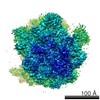 2695MC 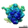 2696C 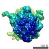 2711C M: map data used to model this data C: citing same article ( |
|---|---|
| Similar structure data |

-Links
 PDBj
PDBj


- Assembly
| Deposited unit | 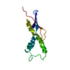
|
|---|---|
| 1 |
|
-Components
| #1: Protein | Mass: 12711.546 Da / Num. of mol.: 1 / Fragment: RIBOSOME BINDING DOMAIN, RESIDUES 1-115 Source method: isolated from a genetically manipulated source Source: (gene. exp.) |
|---|
-Experimental details
-Experiment
| Experiment | Method: ELECTRON MICROSCOPY |
|---|---|
| EM experiment | Aggregation state: PARTICLE / 3D reconstruction method: single particle reconstruction |
- Sample preparation
| Component | Name: TNAC-STALLED-RNC WITH TRIGGER FACTOR / Type: COMPLEX |
|---|---|
| Buffer solution | Name: 20 MM HEPES, 100 MM KOAC, 10 MM MG(OAC)2, 2 MM DTT / pH: 7.4 / Details: 20 MM HEPES, 100 MM KOAC, 10 MM MG(OAC)2, 2 MM DTT |
| Specimen | Embedding applied: NO / Shadowing applied: NO / Staining applied: NO / Vitrification applied: YES |
| Specimen support | Details: HOLEY CARBON |
| Vitrification | Instrument: FEI VITROBOT MARK III / Cryogen name: ETHANE Details: VITRIFICATION 1 -- CRYOGEN- ETHANE, HUMIDITY- 100, INSTRUMENT- FEI VITROBOT MARK III, METHOD- BLOT FOR 10 SECONDS BEFORE PLUNGING, USE 2 LAYERS OF FILTER PAPER V, |
- Electron microscopy imaging
| Experimental equipment |  Model: Tecnai F30 / Image courtesy: FEI Company |
|---|---|
| Microscopy | Model: FEI TECNAI F30 / Date: Nov 21, 2010 |
| Electron gun | Electron source:  FIELD EMISSION GUN / Accelerating voltage: 300 kV / Illumination mode: FLOOD BEAM FIELD EMISSION GUN / Accelerating voltage: 300 kV / Illumination mode: FLOOD BEAM |
| Electron lens | Mode: BRIGHT FIELD / Nominal magnification: 39000 X / Calibrated magnification: 38900 X / Nominal defocus max: 3500 nm / Nominal defocus min: 1200 nm / Cs: 2 mm |
| Image recording | Electron dose: 20 e/Å2 / Film or detector model: KODAK SO-163 FILM |
| Image scans | Num. digital images: 221 |
- Processing
| EM software | Name: SPIDER / Category: 3D reconstruction | ||||||||||||
|---|---|---|---|---|---|---|---|---|---|---|---|---|---|
| CTF correction | Details: ON VOLUMES (SPIDER) | ||||||||||||
| Symmetry | Point symmetry: C1 (asymmetric) | ||||||||||||
| 3D reconstruction | Method: PROJECTION MATCHING / Resolution: 7.7 Å / Num. of particles: 100931 Details: SUBMISSION BASED ON EXPERIMENTAL DATA FROM EMDB EMD-2695. (DEPOSITION ID: 12641). Symmetry type: POINT | ||||||||||||
| Refinement | Highest resolution: 7.7 Å | ||||||||||||
| Refinement step | Cycle: LAST / Highest resolution: 7.7 Å
|
