 Movie
Movie Controller

[English] 日本語
 Yorodumi
- EMDB-8423: High affinity anchoring of the decoration protein pb10 onto the b... -
+ Open data

- Basic information
| Entry | Database: EMDB / ID: EMD-8423 | |||||||||
|---|---|---|---|---|---|---|---|---|---|---|
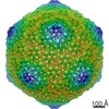
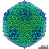
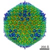
| Title | High affinity anchoring of the decoration protein pb10 onto the bacteriophage T5 capsid | |||||||||
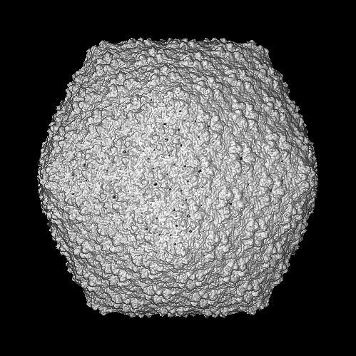
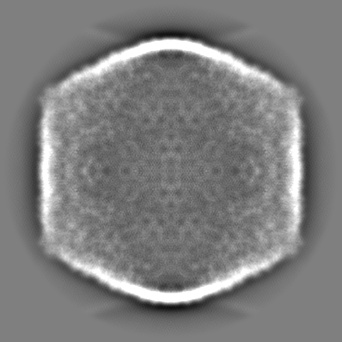
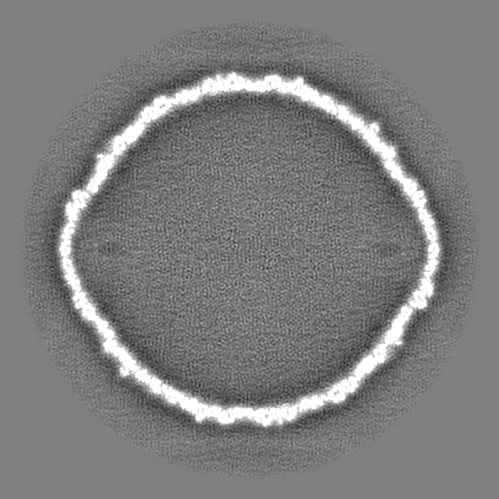
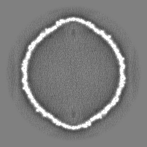
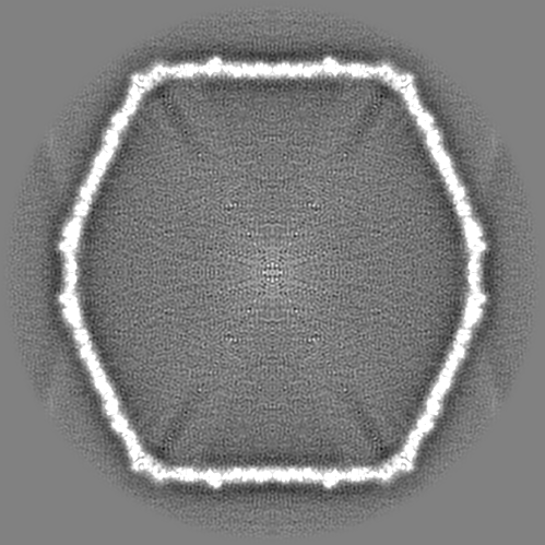
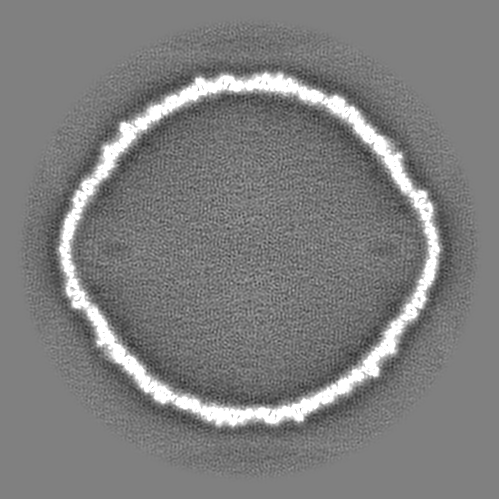
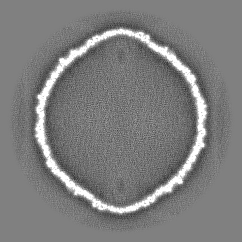
 Map data Map data | Empty expanded capsid of bacteriophage T5 without the decoration protein | |||||||||
 Sample Sample |
| |||||||||
| Biological species |  Enterobacteria phage T5 (virus) Enterobacteria phage T5 (virus) | |||||||||
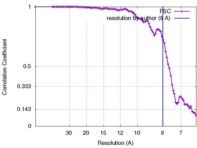
| Method | single particle reconstruction / cryo EM / Resolution: 8.0 Å | |||||||||
 Authors Authors | Conway J / Huet A | |||||||||
 Citation Citation | Journal: Sci Rep / Year: 2017 Title: High affinity anchoring of the decoration protein pb10 onto the bacteriophage T5 capsid. Authors: Emeline Vernhes / Madalena Renouard / Bernard Gilquin / Philippe Cuniasse / Dominique Durand / Patrick England / Sylviane Hoos / Alexis Huet / James F Conway / Anatoly Glukhov / Vladimir ...Authors: Emeline Vernhes / Madalena Renouard / Bernard Gilquin / Philippe Cuniasse / Dominique Durand / Patrick England / Sylviane Hoos / Alexis Huet / James F Conway / Anatoly Glukhov / Vladimir Ksenzenko / Eric Jacquet / Naïma Nhiri / Sophie Zinn-Justin / Pascale Boulanger /    Abstract: Bacteriophage capsids constitute icosahedral shells of exceptional stability that protect the viral genome. Many capsids display on their surface decoration proteins whose structure and function ...Bacteriophage capsids constitute icosahedral shells of exceptional stability that protect the viral genome. Many capsids display on their surface decoration proteins whose structure and function remain largely unknown. The decoration protein pb10 of phage T5 binds at the centre of the 120 hexamers formed by the major capsid protein. Here we determined the 3D structure of pb10 and investigated its capsid-binding properties using NMR, SAXS, cryoEM and SPR. Pb10 consists of an α-helical capsid-binding domain and an Ig-like domain exposed to the solvent. It binds to the T5 capsid with a remarkably high affinity and its binding kinetics is characterized by a very slow dissociation rate. We propose that the conformational exchange events observed in the capsid-binding domain enable rearrangements upon binding that contribute to the quasi-irreversibility of the pb10-capsid interaction. Moreover we show that pb10 binding is a highly cooperative process, which favours immediate rebinding of newly dissociated pb10 to the 120 hexamers of the capsid protein. In extreme conditions, pb10 protects the phage from releasing its genome. We conclude that pb10 may function to reinforce the capsid thus favouring phage survival in harsh environments. | |||||||||
| History |
|
- Structure visualization
| Movie |
 Movie viewer Movie viewer |
|---|---|
| Structure viewer | EM map: SurfViewMolmilJmol/JSmol |
| Supplemental images |

- Downloads & links
-EMDB archive
| Map data | emd_8423.map.gz | 172.7 MB | EMDB map data format | |
|---|---|---|---|---|
| Header (meta data) | emd-8423-v30.xmlemd-8423.xml | 11.4 KB 11.4 KB | Display Display | EMDB header |
| FSC (resolution estimation) | emd_8423_fsc.xml | 8.1 KB | Display | FSC data file |
| Images | 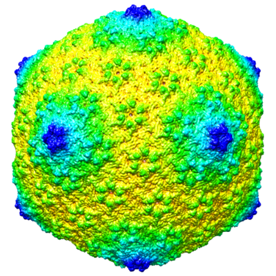 emd_8423.png emd_8423.png | 288.2 KB | ||
| Archive directory |  http://ftp.pdbj.org/pub/emdb/structures/EMD-8423ftp://ftp.pdbj.org/pub/emdb/structures/EMD-8423 http://ftp.pdbj.org/pub/emdb/structures/EMD-8423ftp://ftp.pdbj.org/pub/emdb/structures/EMD-8423 | HTTPS FTP |
-Validation report
| Summary document | emd_8423_validation.pdf.gz | 78.3 KB | Display | EMDB validaton report |
|---|---|---|---|---|
| Full document | emd_8423_full_validation.pdf.gz | 77.4 KB | Display | |
| Data in XML | emd_8423_validation.xml.gz | 494 B | Display | |
| Arichive directory | https://ftp.pdbj.org/pub/emdb/validation_reports/EMD-8423ftp://ftp.pdbj.org/pub/emdb/validation_reports/EMD-8423 | HTTPS FTP |
-Related structure data


-Links
| EMDB pages | EMDB (EBI/PDBe) / EMDataResource |
|---|
-Map
| File | Download / File: emd_8423.map.gz / Format: CCP4 / Size: 474 MB / Type: IMAGE STORED AS FLOATING POINT NUMBER (4 BYTES) | ||||||||||||||||||||||||||||||||||||||||||||||||||||||||||||||||||||
|---|---|---|---|---|---|---|---|---|---|---|---|---|---|---|---|---|---|---|---|---|---|---|---|---|---|---|---|---|---|---|---|---|---|---|---|---|---|---|---|---|---|---|---|---|---|---|---|---|---|---|---|---|---|---|---|---|---|---|---|---|---|---|---|---|---|---|---|---|---|
| Annotation | Empty expanded capsid of bacteriophage T5 without the decoration protein | ||||||||||||||||||||||||||||||||||||||||||||||||||||||||||||||||||||
| Projections & slices | Image control
Images are generated by Spider. | ||||||||||||||||||||||||||||||||||||||||||||||||||||||||||||||||||||
| Voxel size | X=Y=Z: 2.14 Å | ||||||||||||||||||||||||||||||||||||||||||||||||||||||||||||||||||||
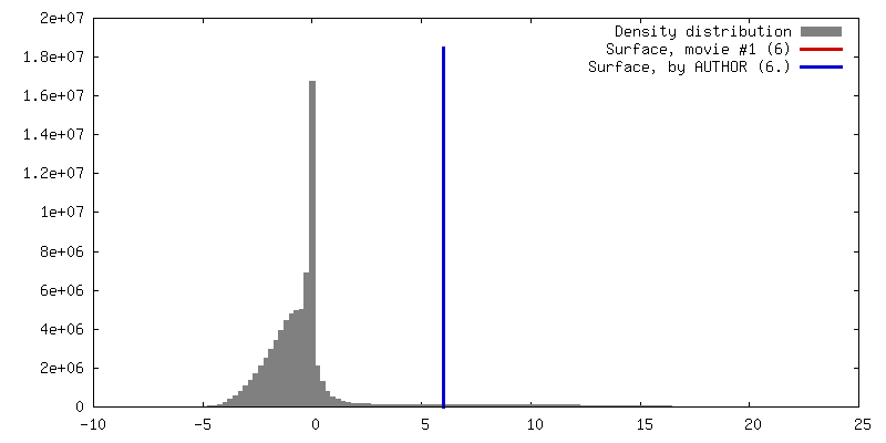
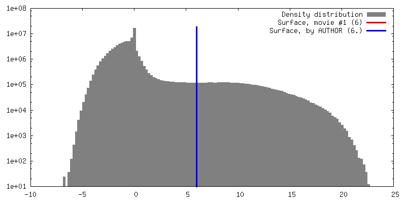
| Density |
| ||||||||||||||||||||||||||||||||||||||||||||||||||||||||||||||||||||
| Symmetry | Space group: 1 | ||||||||||||||||||||||||||||||||||||||||||||||||||||||||||||||||||||
| Details | EMDB XML:
CCP4 map header:
| ||||||||||||||||||||||||||||||||||||||||||||||||||||||||||||||||||||
 Z (Sec.)
 Y (Row.)
 X (Col.)


-Supplemental data
- Sample components
-Entire : Enterobacteria phage T5
| Entire | Name: Enterobacteria phage T5 (virus) |
|---|---|
| Components |
|
-Supramolecule #1: Enterobacteria phage T5
| Supramolecule | Name: Enterobacteria phage T5 / type: virus / ID: 1 / Parent: 0 / Macromolecule list: #1 Details: from AMN5 amber mutant strain - mutation in the terminase. NCBI-ID: 10726 / Sci species name: Enterobacteria phage T5 / Virus type: VIRION / Virus isolate: OTHER / Virus enveloped: No / Virus empty: Yes |
|---|---|
| Host (natural) | Organism: |
| Virus shell | Shell ID: 1 / Name: capsid / Diameter: 900.0 Å / T number (triangulation number): 13 |
-Experimental details
-Structure determination
| Method | cryo EM |
|---|---|
 Processing Processing | single particle reconstruction |
| Aggregation state | particle |
-Sample preparation
| Concentration | 1 mg/mL |
|---|---|
| Buffer | pH: 7.6 / Component - Concentration: 10.0 mM / Component - Name: tris |
| Grid | Material: COPPER / Mesh: 400 / Pretreatment - Type: GLOW DISCHARGE |
| Vitrification | Cryogen name: ETHANE-PROPANE / Chamber humidity: 90 % / Chamber temperature: 293 K / Instrument: FEI VITROBOT MARK II |
- Electron microscopy
| Microscope | FEI TITAN KRIOS |
|---|---|
| Image recording | Film or detector model: FEI FALCON II (4k x 4k) / Digitization - Sampling interval: 14.0 µm / Number real images: 5171 / Average exposure time: 2.0 sec. / Average electron dose: 30.0 e/Å2 |
| Electron beam | Acceleration voltage: 300 kV / Electron source:  FIELD EMISSION GUN FIELD EMISSION GUN |
| Electron optics | Illumination mode: FLOOD BEAM / Imaging mode: BRIGHT FIELD / Cs: 2.7 mm / Nominal defocus max: 5.0 µm / Nominal defocus min: 1.0 µm / Nominal magnification: 75000 |
| Sample stage | Specimen holder model: FEI TITAN KRIOS AUTOGRID HOLDER / Cooling holder cryogen: NITROGEN |
| Experimental equipment |  Model: Titan Krios / Image courtesy: FEI Company |
+Image processing
-Atomic model buiding 1
| Refinement | Space: REAL / Protocol: FLEXIBLE FIT |
|---|
