[histone H3]-lysine79 N-trimethyltransferase / histone H3K79 methyltransferase activity / histone H3K79 trimethyltransferase activity / regulation of transcription regulatory region DNA binding / hypothalamus gonadotrophin-releasing hormone neuron development / female meiosis I / positive regulation of protein monoubiquitination / regulation of receptor signaling pathway via JAK-STAT / fat pad development / mitochondrion transport along microtubule ...[histone H3]-lysine79 N-trimethyltransferase / histone H3K79 methyltransferase activity / histone H3K79 trimethyltransferase activity / regulation of transcription regulatory region DNA binding / hypothalamus gonadotrophin-releasing hormone neuron development / female meiosis I / positive regulation of protein monoubiquitination / regulation of receptor signaling pathway via JAK-STAT / fat pad development / mitochondrion transport along microtubule / histone H3 methyltransferase activity / histone methyltransferase activity / female gonad development / seminiferous tubule development / male meiosis I / positive regulation of intrinsic apoptotic signaling pathway by p53 class mediator / subtelomeric heterochromatin formation / energy homeostasis / neuron projection morphogenesis / regulation of neuron apoptotic process / regulation of proteasomal protein catabolic process / Maturation of protein E / Maturation of protein E / ER Quality Control Compartment (ERQC) / Myoclonic epilepsy of Lafora / FLT3 signaling by CBL mutants / IRAK2 mediated activation of TAK1 complex / Prevention of phagosomal-lysosomal fusion / Alpha-protein kinase 1 signaling pathway / Glycogen synthesis / IRAK1 recruits IKK complex / IRAK1 recruits IKK complex upon TLR7/8 or 9 stimulation / Endosomal Sorting Complex Required For Transport (ESCRT) / Membrane binding and targetting of GAG proteins / Negative regulation of FLT3 / Regulation of TBK1, IKKε (IKBKE)-mediated activation of IRF3, IRF7 / PTK6 Regulates RTKs and Their Effectors AKT1 and DOK1 / Regulation of TBK1, IKKε-mediated activation of IRF3, IRF7 upon TLR3 ligation / Constitutive Signaling by NOTCH1 HD Domain Mutants / IRAK2 mediated activation of TAK1 complex upon TLR7/8 or 9 stimulation / NOTCH2 Activation and Transmission of Signal to the Nucleus / TICAM1,TRAF6-dependent induction of TAK1 complex / telomere organization / TICAM1-dependent activation of IRF3/IRF7 / APC/C:Cdc20 mediated degradation of Cyclin B / Downregulation of ERBB4 signaling / Regulation of FZD by ubiquitination / APC-Cdc20 mediated degradation of Nek2A / p75NTR recruits signalling complexes / InlA-mediated entry of Listeria monocytogenes into host cells / TRAF6 mediated IRF7 activation in TLR7/8 or 9 signaling / TRAF6-mediated induction of TAK1 complex within TLR4 complex / Regulation of pyruvate metabolism / NF-kB is activated and signals survival / Regulation of innate immune responses to cytosolic DNA / Pexophagy / Downregulation of ERBB2:ERBB3 signaling / NRIF signals cell death from the nucleus / Regulation of PTEN localization / DNA damage checkpoint signaling / VLDLR internalisation and degradation / Activated NOTCH1 Transmits Signal to the Nucleus / Regulation of BACH1 activity / Synthesis of active ubiquitin: roles of E1 and E2 enzymes / MAP3K8 (TPL2)-dependent MAPK1/3 activation / Translesion synthesis by REV1 / TICAM1, RIP1-mediated IKK complex recruitment / Translesion synthesis by POLK / InlB-mediated entry of Listeria monocytogenes into host cell / positive regulation of protein ubiquitination / Activation of IRF3, IRF7 mediated by TBK1, IKKε (IKBKE) / JNK (c-Jun kinases) phosphorylation and activation mediated by activated human TAK1 / Josephin domain DUBs / Downregulation of TGF-beta receptor signaling / Translesion synthesis by POLI / IKK complex recruitment mediated by RIP1 / Gap-filling DNA repair synthesis and ligation in GG-NER / Regulation of activated PAK-2p34 by proteasome mediated degradation / PINK1-PRKN Mediated Mitophagy / TGF-beta receptor signaling in EMT (epithelial to mesenchymal transition) / regulation of mitochondrial membrane potential / TNFR1-induced NF-kappa-B signaling pathway / Autodegradation of Cdh1 by Cdh1:APC/C / TCF dependent signaling in response to WNT / APC/C:Cdc20 mediated degradation of Securin / Regulation of NF-kappa B signaling / N-glycan trimming in the ER and Calnexin/Calreticulin cycle / activated TAK1 mediates p38 MAPK activation / Asymmetric localization of PCP proteins / Ubiquitin-dependent degradation of Cyclin D / SCF-beta-TrCP mediated degradation of Emi1 / NIK-->noncanonical NF-kB signaling / TNFR2 non-canonical NF-kB pathway / Regulation of signaling by CBL / AUF1 (hnRNP D0) binds and destabilizes mRNA / NOTCH3 Activation and Transmission of Signal to the Nucleus / Negative regulators of DDX58/IFIH1 signaling / Vpu mediated degradation of CD4 / Negative regulation of FGFR3 signaling / Assembly of the pre-replicative complex Similarity search - Function
National Institutes of Health/National Institute of General Medical Sciences (NIH/NIGMS)
GM095822
United States
Citation
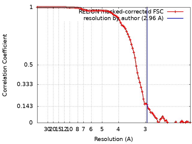
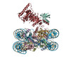
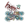
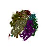
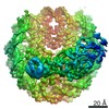
Journal: Cell / Year: 2019 Title: Mechanism of Cross-talk between H2B Ubiquitination and H3 Methylation by Dot1L. Authors: Evan J Worden / Niklas A Hoffmann / Chad W Hicks / Cynthia Wolberger / Abstract: Methylation of histone H3 K79 by Dot1L is a hallmark of actively transcribed genes that depends on monoubiquitination of H2B K120 (H2B-Ub) and is an example of histone modification cross-talk that is ...Methylation of histone H3 K79 by Dot1L is a hallmark of actively transcribed genes that depends on monoubiquitination of H2B K120 (H2B-Ub) and is an example of histone modification cross-talk that is conserved from yeast to humans. We report here cryo-EM structures of Dot1L bound to ubiquitinated nucleosome that show how H2B-Ub stimulates Dot1L activity and reveal a role for the histone H4 tail in positioning Dot1L. We find that contacts mediated by Dot1L and the H4 tail induce a conformational change in the globular core of histone H3 that reorients K79 from an inaccessible position, thus enabling this side chain to insert into the active site in a position primed for catalysis. Our study provides a comprehensive mechanism of cross-talk between histone ubiquitination and methylation and reveals structural plasticity in histones that makes it possible for histone-modifying enzymes to access residues within the nucleosome core.
History
Deposition
Jan 2, 2019
-
Header (metadata) release
Jan 23, 2019
-
Map release
Feb 20, 2019
-
Update
Oct 16, 2024
-
Current status
Oct 16, 2024
Processing site: RCSB / Status: Released
-
Structure visualization
Movie
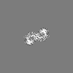
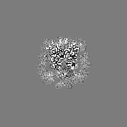
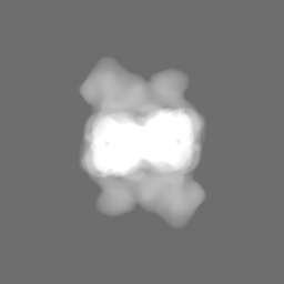
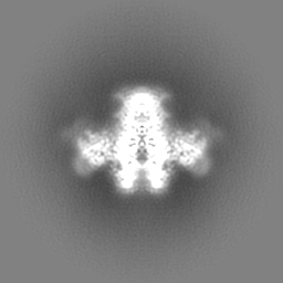
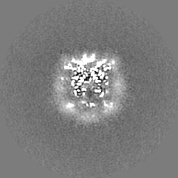
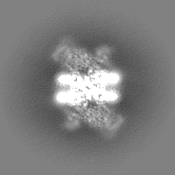
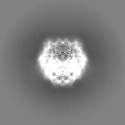
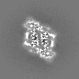
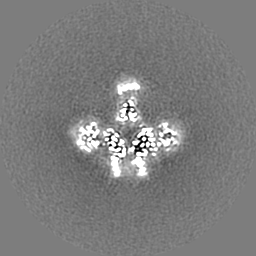
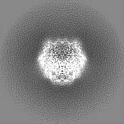
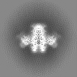
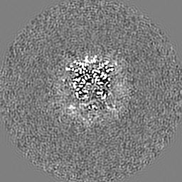
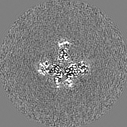
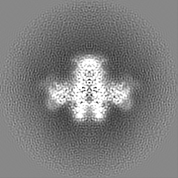
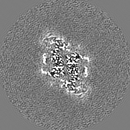
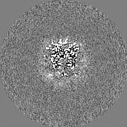
Surface view with section colored by density value
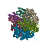
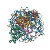
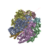
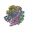
Entire : Active state Dot1L in complex with the H2B-Ub nucleosome
Entire
Name: Active state Dot1L in complex with the H2B-Ub nucleosome
Components
Complex: Active state Dot1L in complex with the H2B-Ub nucleosome
Complex: Dot1L
Protein or peptide: Histone-lysine N-methyltransferase, H3 lysine-79 specific
Complex: H2H2B-Ub nucleosome
Complex: histone core
Protein or peptide: Histone H3.2
Protein or peptide: Histone H4
Protein or peptide: Histone H2A type 1
Protein or peptide: Histone H2B 1.1
Complex: ubiquitin
Protein or peptide: Ubiquitin
Complex: DNA
DNA: 601 DNA Strand 1
DNA: 601 DNA Strand 2
Ligand: S-ADENOSYLMETHIONINE
+
Supramolecule #1: Active state Dot1L in complex with the H2B-Ub nucleosome
Supramolecule
Name: Active state Dot1L in complex with the H2B-Ub nucleosome type: complex / ID: 1 / Parent: 0 / Macromolecule list: #1-#8 / Details: 2:1 complex of Dot1L bound to the nucleosome
Details: Solutions were prepared on the day of freezing and filtered though a 0.2 um filter prior to use.
Grid
Model: C-flat-2/2 / Material: COPPER / Mesh: 400 / Support film - Material: CARBON / Support film - topology: HOLEY ARRAY / Pretreatment - Type: GLOW DISCHARGE / Pretreatment - Time: 30 sec. / Pretreatment - Atmosphere: AIR
Vitrification
Cryogen name: ETHANE / Chamber humidity: 100 % / Chamber temperature: 277 K / Instrument: FEI VITROBOT MARK IV / Details: Blot once for 3.5 seconds before freezing..
Details
Crosslinked with glutaraldehyde
-
Electron microscopy
Microscope
FEI TITAN KRIOS
Image recording
Film or detector model: GATAN K2 SUMMIT (4k x 4k) / Detector mode: SUPER-RESOLUTION / Digitization - Frames/image: 1-40 / Number grids imaged: 2 / Number real images: 2284 / Average exposure time: 8.2 sec. / Average electron dose: 50.0 e/Å2 / Details: 3 exposures per hole
Electron beam
Acceleration voltage: 300 kV / Electron source: FIELD EMISSION GUN
In the structure databanks used in Yorodumi, some data are registered as the other names, "COVID-19 virus" and "2019-nCoV". Here are the details of the virus and the list of structure data.
Jan 31, 2019. EMDB accession codes are about to change! (news from PDBe EMDB page)
EMDB accession codes are about to change! (news from PDBe EMDB page)
The allocation of 4 digits for EMDB accession codes will soon come to an end. Whilst these codes will remain in use, new EMDB accession codes will include an additional digit and will expand incrementally as the available range of codes is exhausted. The current 4-digit format prefixed with “EMD-” (i.e. EMD-XXXX) will advance to a 5-digit format (i.e. EMD-XXXXX), and so on. It is currently estimated that the 4-digit codes will be depleted around Spring 2019, at which point the 5-digit format will come into force.
The EM Navigator/Yorodumi systems omit the EMD- prefix.
Related info.:Q: What is EMD? / ID/Accession-code notation in Yorodumi/EM Navigator
Yorodumi is a browser for structure data from EMDB, PDB, SASBDB, etc.
This page is also the successor to EM Navigator detail page, and also detail information page/front-end page for Omokage search.
The word "yorodu" (or yorozu) is an old Japanese word meaning "ten thousand". "mi" (miru) is to see.
Related info.:EMDB / PDB / SASBDB / Comparison of 3 databanks / Yorodumi Search / Aug 31, 2016. New EM Navigator & Yorodumi / Yorodumi Papers / Jmol/JSmol / Function and homology information / Changes in new EM Navigator and Yorodumi
 Movie
Movie Controller

 Yorodumi
 Open data
 Basic information
 Map data
 Sample
 Keywords
 Function and homology information
 Homo sapiens (human) /
  Authors
 United States, 1 items
  Citation
 Structure visualization
 Downloads & links
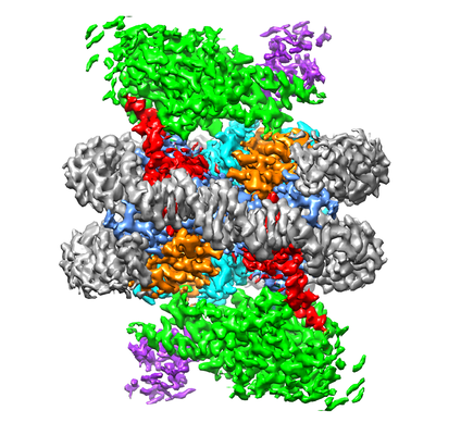 emd_9384.png
 http://ftp.pdbj.org/pub/emdb/structures/EMD-9384


 Z (Sec.)
 Y (Row.)
 X (Col.)


 Sample components

 Processing
 Electron microscopy
 FIELD EMISSION GUN