Movie
Movie Controller


+ Open data

- Basic information
| Entry | Database: EMDB / ID: EMD-8711 | |||||||||||||||
|---|---|---|---|---|---|---|---|---|---|---|---|---|---|---|---|---|
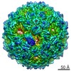
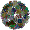
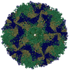
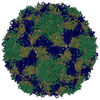
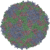
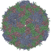
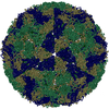
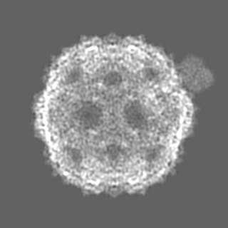
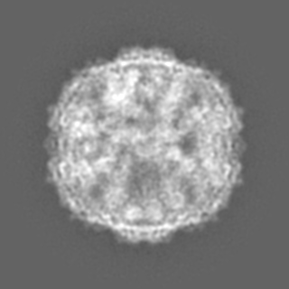
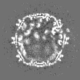
| Title | Phage Qbeta in complex with MurA | |||||||||||||||
 Map data Map data | Enterobacteria phage Qbeta virion with MurA | |||||||||||||||
 Sample Sample |
| |||||||||||||||
 Keywords Keywords | Qbeta / ssRNA / phage / virus / VIRUS-TRANSFERASE complex | |||||||||||||||
| Function / homology |  Function and homology information Function and homology informationsymbiont-mediated suppression of host cell wall biogenesis / symbiont-mediated suppression of host peptidoglycan biosynthetic process / viral release via suppression of host peptidoglycan biosynthetic process / virion attachment to host cell pilus / UDP-N-acetylglucosamine 1-carboxyvinyltransferase activity / UDP-N-acetylgalactosamine biosynthetic process / UDP-N-acetylglucosamine 1-carboxyvinyltransferase / adhesion receptor-mediated virion attachment to host cell / peptidoglycan biosynthetic process / viral release from host cell by cytolysis ...symbiont-mediated suppression of host cell wall biogenesis / symbiont-mediated suppression of host peptidoglycan biosynthetic process / viral release via suppression of host peptidoglycan biosynthetic process / virion attachment to host cell pilus / UDP-N-acetylglucosamine 1-carboxyvinyltransferase activity / UDP-N-acetylgalactosamine biosynthetic process / UDP-N-acetylglucosamine 1-carboxyvinyltransferase / adhesion receptor-mediated virion attachment to host cell / peptidoglycan biosynthetic process / viral release from host cell by cytolysis / cell wall organization / virion component / regulation of cell shape / cell division / cytoplasm Similarity search - Function | |||||||||||||||
| Biological species |   Escherichia phage Qbeta (virus) / Enterobacteria phage Qbeta (virus) Escherichia phage Qbeta (virus) / Enterobacteria phage Qbeta (virus) | |||||||||||||||
| Method | single particle reconstruction / cryo EM / Resolution: 5.7 Å | |||||||||||||||
 Authors Authors | Cui Z / Zhang J | |||||||||||||||
| Funding support |  United States, 4 items United States, 4 items
| |||||||||||||||
 Citation Citation | Journal: Proc Natl Acad Sci U S A / Year: 2017 Title: Structures of Qβ virions, virus-like particles, and the Qβ-MurA complex reveal internal coat proteins and the mechanism of host lysis. Authors: Zhicheng Cui / Karl V Gorzelnik / Jeng-Yih Chang / Carrie Langlais / Joanita Jakana / Ry Young / Junjie Zhang / Abstract: In single-stranded RNA bacteriophages (ssRNA phages) a single copy of the maturation protein binds the genomic RNA (gRNA) and is required for attachment of the phage to the host pilus. For the ...In single-stranded RNA bacteriophages (ssRNA phages) a single copy of the maturation protein binds the genomic RNA (gRNA) and is required for attachment of the phage to the host pilus. For the canonical Qβ the maturation protein, A, has an additional role as the lysis protein, by its ability to bind and inhibit MurA, which is involved in peptidoglycan biosynthesis. Here, we determined structures of Qβ virions, virus-like particles, and the Qβ-MurA complex using single-particle cryoelectron microscopy, at 4.7-Å, 3.3-Å, and 6.1-Å resolutions, respectively. We identified the outer surface of the β-region in A as the MurA-binding interface. Moreover, the pattern of MurA mutations that block Qβ lysis and the conformational changes of MurA that facilitate A binding were found to be due to the intimate fit between A and the region encompassing the closed catalytic cleft of substrate-liganded MurA. Additionally, by comparing the Qβ virion with Qβ virus-like particles that lack a maturation protein, we observed a structural rearrangement in the capsid coat proteins that is required to package the viral gRNA in its dominant conformation. Unexpectedly, we found a coat protein dimer sequestered in the interior of the virion. This coat protein dimer binds to the gRNA and interacts with the buried α-region of A, suggesting that it is sequestered during the early stage of capsid formation to promote the gRNA condensation required for genome packaging. These internalized coat proteins are the most asymmetrically arranged major capsid proteins yet observed in virus structures. | |||||||||||||||
| History |
|
- Structure visualization
| Movie |
Movie viewer |
|---|---|
| Structure viewer | EM map: SurfViewMolmilJmol/JSmol |
| Supplemental images |

- Downloads & links
-EMDB archive
| Map data | emd_8711.map.gz | 15.4 MB | EMDB map data format | |
|---|---|---|---|---|
| Header (meta data) | emd-8711-v30.xmlemd-8711.xml | 17.7 KB 17.7 KB | Display Display | EMDB header |
| Images | 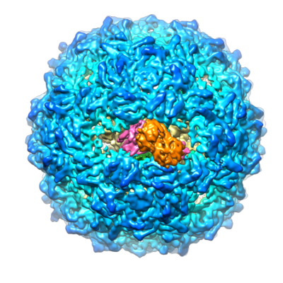 emd_8711.png emd_8711.png | 221.4 KB | ||
| Filedesc metadata | emd-8711.cif.gz | 5.9 KB | ||
| Others | emd_8711_additional.map.gz | 41.5 MB | ||
| Archive directory |  http://ftp.pdbj.org/pub/emdb/structures/EMD-8711ftp://ftp.pdbj.org/pub/emdb/structures/EMD-8711 http://ftp.pdbj.org/pub/emdb/structures/EMD-8711ftp://ftp.pdbj.org/pub/emdb/structures/EMD-8711 | HTTPS FTP |
-Related structure data
| Related structure data |  5vm7MC  8707C  8708C 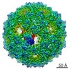 8709C  8710C  5vlyC  5vlzC C: citing same article ( M: atomic model generated by this map |
|---|---|
| Similar structure data |


-Links
| EMDB pages | EMDB (EBI/PDBe) / EMDataResource |
|---|---|
| Related items in Molecule of the Month |

-Map
| File | Download / File: emd_8711.map.gz / Format: CCP4 / Size: 125 MB / Type: IMAGE STORED AS FLOATING POINT NUMBER (4 BYTES) | ||||||||||||||||||||||||||||||||||||||||||||||||||||||||||||
|---|---|---|---|---|---|---|---|---|---|---|---|---|---|---|---|---|---|---|---|---|---|---|---|---|---|---|---|---|---|---|---|---|---|---|---|---|---|---|---|---|---|---|---|---|---|---|---|---|---|---|---|---|---|---|---|---|---|---|---|---|---|
| Annotation | Enterobacteria phage Qbeta virion with MurA | ||||||||||||||||||||||||||||||||||||||||||||||||||||||||||||
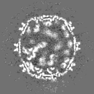
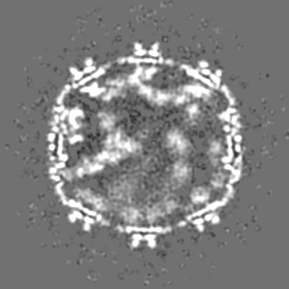
| Projections & slices | Image control
Images are generated by Spider. | ||||||||||||||||||||||||||||||||||||||||||||||||||||||||||||
| Voxel size | X=Y=Z: 1.25 Å | ||||||||||||||||||||||||||||||||||||||||||||||||||||||||||||
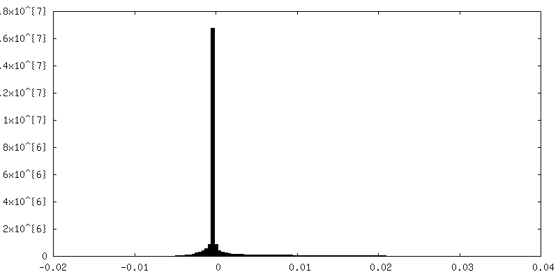
| Density |
| ||||||||||||||||||||||||||||||||||||||||||||||||||||||||||||
| Symmetry | Space group: 1 | ||||||||||||||||||||||||||||||||||||||||||||||||||||||||||||
| Details | EMDB XML:
CCP4 map header:
| ||||||||||||||||||||||||||||||||||||||||||||||||||||||||||||
 Z (Sec.)
 Y (Row.)
 X (Col.)


-Supplemental data
-Additional map: Enterobacteria phage Qbeta virion with MurA, additional map
| File | emd_8711_additional.map | ||||||||||||
|---|---|---|---|---|---|---|---|---|---|---|---|---|---|
| Annotation | Enterobacteria phage Qbeta virion with MurA, additional map | ||||||||||||
| Projections & Slices |
| ||||||||||||
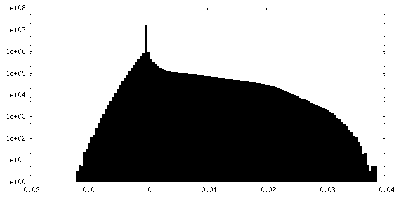
| Density Histograms |

- Sample components
-Entire : Enterobacteria phage Qbeta virion with MurA
| Entire | Name: Enterobacteria phage Qbeta virion with MurA |
|---|---|
| Components |
|
-Supramolecule #1: Enterobacteria phage Qbeta virion with MurA
| Supramolecule | Name: Enterobacteria phage Qbeta virion with MurA / type: complex / ID: 1 / Parent: 0 / Macromolecule list: all |
|---|
-Supramolecule #3: UDP-N-acetylglucosamine 1-carboxyvinyltransferase
| Supramolecule | Name: UDP-N-acetylglucosamine 1-carboxyvinyltransferase / type: complex / ID: 3 / Parent: 1 / Macromolecule list: #2 |
|---|---|
| Source (natural) | Organism: |
-Supramolecule #2: Enterobacteria phage Qbeta
| Supramolecule | Name: Enterobacteria phage Qbeta / type: virus / ID: 2 / Parent: 1 / Macromolecule list: #1 / NCBI-ID: 39803 / Sci species name: Enterobacteria phage Qbeta / Virus type: VIRION / Virus isolate: SPECIES / Virus enveloped: No / Virus empty: No |
|---|
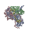
-Macromolecule #1: Maturation protein A2
| Macromolecule | Name: Maturation protein A2 / type: protein_or_peptide / ID: 1 / Number of copies: 1 / Enantiomer: LEVO |
|---|---|
| Source (natural) | Organism: Escherichia phage Qbeta (virus) |
| Molecular weight | Theoretical: 48.613078 KDa |
| Sequence | String: MPKLPRGLRF GADNEILNDF QELWFPDLFI ESSDTHPWYT LKGRVLNAHL DDRLPNVGGR QVRRTPHRVT VPIASSGLRP VTTVQYDPA ALSFLLNARV DWDFGNGDSA NLVINDFLFR TFAPKEFDFS NSLVPRYTQA FSAFNAKYGT MIGEGLETIK Y LGLLLRRL ...String: MPKLPRGLRF GADNEILNDF QELWFPDLFI ESSDTHPWYT LKGRVLNAHL DDRLPNVGGR QVRRTPHRVT VPIASSGLRP VTTVQYDPA ALSFLLNARV DWDFGNGDSA NLVINDFLFR TFAPKEFDFS NSLVPRYTQA FSAFNAKYGT MIGEGLETIK Y LGLLLRRL REGYRAVKRG DLRALRRVIQ SYHNGKWKPA TAGNLWLEFR YGLMPLFYDI RDVMLDWQNR HDKIQRLLRF SV GHGEDYV VEFDNLYPAV AYFKLKGEIT LERRHRHGIS YANREGYAVF DNGSLRPVSD WKELATAFIN PHEVAWELTP YSF VVDWFL NVGDILAQQG QLYHNIDIVD GFDRRDIRLK SFTIKGERNG RPVNVSASLS AVDLFYSRLH TSNLPFATLD LDTT FSSFK HVLDSIFLLT QRVKR UniProtKB: Maturation protein A2 |
-Macromolecule #2: UDP-N-acetylglucosamine 1-carboxyvinyltransferase
| Macromolecule | Name: UDP-N-acetylglucosamine 1-carboxyvinyltransferase / type: protein_or_peptide / ID: 2 / Number of copies: 1 / Enantiomer: LEVO EC number: UDP-N-acetylglucosamine 1-carboxyvinyltransferase |
|---|---|
| Source (natural) | Organism: Strain: E24377A / ETEC |
| Molecular weight | Theoretical: 44.871543 KDa |
| Sequence | String: MDKFRVQGPT KLQGEVTISG AKNAALPILF AALLAEEPVE IQNVPKLKDV DTSMKLLSQL GAKVERNGSV HIDARDVNVF CAPYDLVKT MRASIWALGP LVARFGQGQV SLPGGCTIGA RPVDLHISGL EQLGATIKLE EGYVKASVDG RLKGAHIVMD K VSVGATVT ...String: MDKFRVQGPT KLQGEVTISG AKNAALPILF AALLAEEPVE IQNVPKLKDV DTSMKLLSQL GAKVERNGSV HIDARDVNVF CAPYDLVKT MRASIWALGP LVARFGQGQV SLPGGCTIGA RPVDLHISGL EQLGATIKLE EGYVKASVDG RLKGAHIVMD K VSVGATVT IMCAATLAEG TTIIENAARE PEIVDTANFL ITLGAKISGQ GTDRIVIEGV ERLGGGVYRV LPDRIETGTF LV AAAISRG KIICRNAQPD TLDAVLAKLR DAGADIEVGE DWISLDMHGK RPKAVNVRTA PHPAFPTDMQ AQFTLLNLVA EGT GFITET VFENRFMHVP ELSRMGAHAE IESNTVICHG VEKLSGAQVM ATDLRASASL VLAGCIAEGT TVVDRIYHID RGYE RIEDK LRALGANIER VKGE UniProtKB: UDP-N-acetylglucosamine 1-carboxyvinyltransferase |
-Experimental details
-Structure determination
| Method | cryo EM |
|---|---|
 Processing Processing | single particle reconstruction |
| Aggregation state | particle |
-Sample preparation
| Buffer | pH: 7 |
|---|---|
| Grid | Model: C-flat-1.2/1.3 / Material: COPPER / Mesh: 400 / Pretreatment - Type: GLOW DISCHARGE / Pretreatment - Time: 60 sec. |
| Vitrification | Cryogen name: ETHANE / Chamber humidity: 100 % / Chamber temperature: 295 K / Instrument: FEI VITROBOT MARK III Details: Blotted for 6s, Plunged into liquid ethane (FEI VITROBOT MARK III). |
- Electron microscopy #1
| Microscopy ID | 1 |
|---|---|
| Microscope | JEOL 3200FSC |
| Image recording | Image recording ID: 1 / Film or detector model: GATAN K2 SUMMIT (4k x 4k) / Detector mode: SUPER-RESOLUTION / Average exposure time: 0.2 sec. / Average electron dose: 1.0 e/Å2 |
| Electron beam | Acceleration voltage: 300 kV / Electron source:  FIELD EMISSION GUN FIELD EMISSION GUN |
| Electron optics | Illumination mode: FLOOD BEAM / Imaging mode: BRIGHT FIELD / Nominal magnification: 30000 |
| Sample stage | Cooling holder cryogen: NITROGEN |
-Electron microscopy #1~
| Microscopy ID | 1 |
|---|---|
| Microscope | FEI TECNAI F20 |
| Image recording | Image recording ID: 2 / Film or detector model: GATAN K2 SUMMIT (4k x 4k) / Detector mode: SUPER-RESOLUTION / Average exposure time: 0.2 sec. / Average electron dose: 1.1 e/Å2 |
| Electron beam | Acceleration voltage: 200 kV / Electron source: FIELD EMISSION GUN |
| Electron optics | Illumination mode: FLOOD BEAM / Imaging mode: BRIGHT FIELD / Nominal magnification: 29000 |
| Experimental equipment |  Model: Tecnai F20 / Image courtesy: FEI Company |