Movie
Movie Controller

[English] 日本語
 Yorodumi
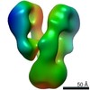
- EMDB-8707: Negative stain reconstruction of Endoplasmic Reticulum HSP40 co-c... -
+ Open data

- Basic information
| Entry | Database: EMDB / ID: EMD-8707 | |||||||||
|---|---|---|---|---|---|---|---|---|---|---|
| Title | Negative stain reconstruction of Endoplasmic Reticulum HSP40 co-chaperone native ERdj3 tetramer. | |||||||||
 Map data Map data | Negative stain reconstruction of native ERdj3 tetramer | |||||||||
 Sample Sample |
| |||||||||
| Function / homology |  Function and homology information Function and homology informationendoplasmic reticulum chaperone complex / XBP1(S) activates chaperone genes / positive regulation of ATP-dependent activity / misfolded protein binding / mRNA modification / protein folding chaperone complex / protein maturation / negative regulation of neurogenesis / : / protein folding ...endoplasmic reticulum chaperone complex / XBP1(S) activates chaperone genes / positive regulation of ATP-dependent activity / misfolded protein binding / mRNA modification / protein folding chaperone complex / protein maturation / negative regulation of neurogenesis / : / protein folding / endoplasmic reticulum lumen / signaling receptor binding / endoplasmic reticulum / : / membrane / nucleus Similarity search - Function | |||||||||
| Biological species |  Homo sapiens (human) Homo sapiens (human) | |||||||||
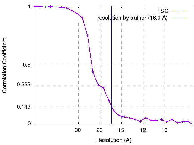
| Method | single particle reconstruction / negative staining / Resolution: 16.9 Å | |||||||||
 Authors Authors | Chowdhury S / Lander GC / Chen K-C / Qu S / Noxon IC / Schonhoft JD / Plate L / Powers ET / Kelly JW / Wiseman RL | |||||||||
 Citation Citation | Journal: Proc Natl Acad Sci U S A / Year: 2017 Title: Structures of Qβ virions, virus-like particles, and the Qβ-MurA complex reveal internal coat proteins and the mechanism of host lysis. Authors: Zhicheng Cui / Karl V Gorzelnik / Jeng-Yih Chang / Carrie Langlais / Joanita Jakana / Ry Young / Junjie Zhang /  Abstract: In single-stranded RNA bacteriophages (ssRNA phages) a single copy of the maturation protein binds the genomic RNA (gRNA) and is required for attachment of the phage to the host pilus. For the ...In single-stranded RNA bacteriophages (ssRNA phages) a single copy of the maturation protein binds the genomic RNA (gRNA) and is required for attachment of the phage to the host pilus. For the canonical Qβ the maturation protein, A, has an additional role as the lysis protein, by its ability to bind and inhibit MurA, which is involved in peptidoglycan biosynthesis. Here, we determined structures of Qβ virions, virus-like particles, and the Qβ-MurA complex using single-particle cryoelectron microscopy, at 4.7-Å, 3.3-Å, and 6.1-Å resolutions, respectively. We identified the outer surface of the β-region in A as the MurA-binding interface. Moreover, the pattern of MurA mutations that block Qβ lysis and the conformational changes of MurA that facilitate A binding were found to be due to the intimate fit between A and the region encompassing the closed catalytic cleft of substrate-liganded MurA. Additionally, by comparing the Qβ virion with Qβ virus-like particles that lack a maturation protein, we observed a structural rearrangement in the capsid coat proteins that is required to package the viral gRNA in its dominant conformation. Unexpectedly, we found a coat protein dimer sequestered in the interior of the virion. This coat protein dimer binds to the gRNA and interacts with the buried α-region of A, suggesting that it is sequestered during the early stage of capsid formation to promote the gRNA condensation required for genome packaging. These internalized coat proteins are the most asymmetrically arranged major capsid proteins yet observed in virus structures. | |||||||||
| History |
|
- Structure visualization
| Movie |
Movie viewer |
|---|---|
| Structure viewer | EM map: SurfViewMolmilJmol/JSmol |
| Supplemental images |
 UCSF Chimera
- Downloads & links
-EMDB archive
| Map data | emd_8707.map.gz | 783.8 KB | EMDB map data format | |
|---|---|---|---|---|
| Header (meta data) | emd-8707-v30.xmlemd-8707.xml | 13.3 KB 13.3 KB | Display Display | EMDB header |
| FSC (resolution estimation) | emd_8707_fsc.xml | 2.6 KB | Display | FSC data file |
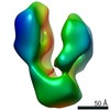
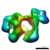
| Images | 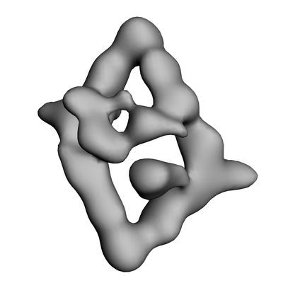 emd_8707.png emd_8707.png | 39.6 KB | ||
| Archive directory |  http://ftp.pdbj.org/pub/emdb/structures/EMD-8707ftp://ftp.pdbj.org/pub/emdb/structures/EMD-8707 http://ftp.pdbj.org/pub/emdb/structures/EMD-8707ftp://ftp.pdbj.org/pub/emdb/structures/EMD-8707 | HTTPS FTP |
-Related structure data
| Related structure data |  8708C 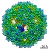 8709C  8710C  8711C  5vlyC  5vlzC  5vm7C C: citing same article ( |
|---|---|
| Similar structure data |

-Links
| EMDB pages | EMDB (EBI/PDBe) / EMDataResource |
|---|---|
| Related items in Molecule of the Month |


-Map
| File | Download / File: emd_8707.map.gz / Format: CCP4 / Size: 844.7 KB / Type: IMAGE STORED AS FLOATING POINT NUMBER (4 BYTES) | ||||||||||||||||||||||||||||||||||||||||||||||||||||||||||||||||||||
|---|---|---|---|---|---|---|---|---|---|---|---|---|---|---|---|---|---|---|---|---|---|---|---|---|---|---|---|---|---|---|---|---|---|---|---|---|---|---|---|---|---|---|---|---|---|---|---|---|---|---|---|---|---|---|---|---|---|---|---|---|---|---|---|---|---|---|---|---|---|
| Annotation | Negative stain reconstruction of native ERdj3 tetramer | ||||||||||||||||||||||||||||||||||||||||||||||||||||||||||||||||||||
| Projections & slices | Image control
Images are generated by Spider. | ||||||||||||||||||||||||||||||||||||||||||||||||||||||||||||||||||||
| Voxel size | X=Y=Z: 4.1 Å | ||||||||||||||||||||||||||||||||||||||||||||||||||||||||||||||||||||
| Density |
| ||||||||||||||||||||||||||||||||||||||||||||||||||||||||||||||||||||
| Symmetry | Space group: 1 | ||||||||||||||||||||||||||||||||||||||||||||||||||||||||||||||||||||
| Details | EMDB XML:
CCP4 map header:
| ||||||||||||||||||||||||||||||||||||||||||||||||||||||||||||||||||||
 Z (Sec.)
 Y (Row.)
 X (Col.)


-Supplemental data
- Sample components
-Entire : Tetrameric complex of native ERdj3 protein complex
| Entire | Name: Tetrameric complex of native ERdj3 protein complex |
|---|---|
| Components |
|
-Supramolecule #1: Tetrameric complex of native ERdj3 protein complex
| Supramolecule | Name: Tetrameric complex of native ERdj3 protein complex / type: complex / ID: 1 / Parent: 0 |
|---|---|
| Source (natural) | Organism: Homo sapiens (human) |
| Recombinant expression | Organism:  |
| Molecular weight | Theoretical: 153 KDa |
-Experimental details
-Structure determination
| Method | negative staining |
|---|---|
 Processing Processing | single particle reconstruction |
| Aggregation state | particle |
-Sample preparation
| Concentration | 0.025 mg/mL |
|---|---|
| Buffer | pH: 7.5 / Details: 50 mM HEPES, 300 mM NaCl, 1% (v/v)Glycerol |
| Staining | Type: NEGATIVE / Material: Uranyl Formate Details: Sample absorbed on carbon surface was stained with 2% (w/v) Uranyl Formate solution, and finally air-dried after blotting off excess stain. |
| Grid | Model: 400 mesh Cu-Rh maxtaform grids (Electron Microscopy Sciences) that were coated with a thin layer of amorphous carbon. Material: COPPER/RHODIUM / Mesh: 400 / Support film - Material: CARBON / Support film - topology: CONTINUOUS / Pretreatment - Type: GLOW DISCHARGE / Pretreatment - Atmosphere: AIR / Details: 15mA current |
| Details | Mono dispersed protein solution |
- Electron microscopy
| Microscope | FEI TECNAI 12 |
|---|---|
| Temperature | Min: 293.0 K / Max: 295.0 K |
| Image recording | Film or detector model: TVIPS TEMCAM-F416 (4k x 4k) / Digitization - Dimensions - Width: 4096 pixel / Digitization - Dimensions - Height: 4096 pixel / Digitization - Sampling interval: 15.6 µm / Number grids imaged: 2 / Number real images: 980 / Average exposure time: 0.79 sec. / Average electron dose: 35.0 e/Å2 Details: Automated image acquisition software Leginon was used for data collection. |
| Electron beam | Acceleration voltage: 120 kV / Electron source: LAB6 |
| Electron optics | Illumination mode: FLOOD BEAM / Imaging mode: BRIGHT FIELD / Cs: 2.2 mm / Nominal defocus max: 1.2 µm / Nominal defocus min: 1.0 µm / Nominal magnification: 52000 |
| Sample stage | Specimen holder model: SIDE ENTRY, EUCENTRIC |