National Institutes of Health/National Institute Of Allergy and Infectious Diseases (NIH/NIAID)
AI095366
米国
引用
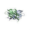
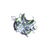
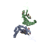
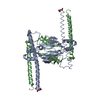
ジャーナル: PLoS Pathog / 年: 2017 タイトル: Assembly, maturation and three-dimensional helical structure of the teratogenic rubella virus. 著者: Vidya Mangala Prasad / Thomas Klose / Michael G Rossmann / 要旨: Viral infections during pregnancy are a significant cause of infant morbidity and mortality. Of these, rubella virus infection is a well-substantiated example that leads to miscarriages or severe ...Viral infections during pregnancy are a significant cause of infant morbidity and mortality. Of these, rubella virus infection is a well-substantiated example that leads to miscarriages or severe fetal defects. However, structural information about the rubella virus has been lacking due to the pleomorphic nature of the virions. Here we report a helical structure of rubella virions using cryo-electron tomography. Sub-tomogram averaging of the surface spikes established the relative positions of the viral glycoproteins, which differed from the earlier icosahedral models of the virus. Tomographic analyses of in vitro assembled nucleocapsids and virions provide a template for viral assembly. Comparisons of immature and mature virions show large rearrangements in the glycoproteins that may be essential for forming the infectious virions. These results present the first known example of a helical membrane-enveloped virus, while also providing a structural basis for its assembly and maturation pathway.
 ムービー
ムービー コントローラー


 データを開く
 基本情報
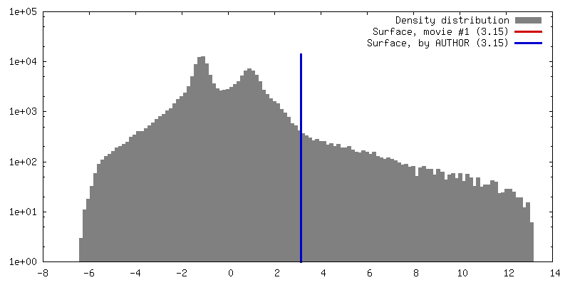
基本情報 マップデータ
 試料
試料 キーワード
キーワード 機能・相同性情報
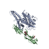
 Rubella virus (風疹ウイルス)
 データ登録者
 米国, 1件
  引用
引用 構造の表示
構造の表示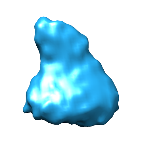
 ダウンロードとリンク
 emd_8248.png
 http://ftp.pdbj.org/pub/emdb/structures/EMD-8248


 Z (Sec.)
 Y (Row.)
 X (Col.)


 試料の構成要素
 Cercopithecus aethiops (ミドリザル)
 解析
解析 電子顕微鏡法
 FIELD EMISSION GUN