[English] 日本語
 Yorodumi
- PDB-1nug: Role of Calcium Ions in the Activation and Activity of the Transg... -
+ Open data
- Basic information
| Entry | Database: PDB / ID: 1nug | ||||||
|---|---|---|---|---|---|---|---|
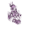
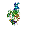
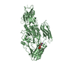
| Title | Role of Calcium Ions in the Activation and Activity of the Transglutaminase 3 Enzyme (2 calciums, 1 Mg, inactive form) | ||||||
 Components Components | Protein-glutamine glutamyltransferase E | ||||||
 Keywords Keywords | TRANSFERASE / Transglutaminase 3 / metalloenzyme / calcium ion | ||||||
| Function / homology |  Function and homology information Function and homology informationprotein-glutamine gamma-glutamyltransferase / protein-glutamine gamma-glutamyltransferase activity / peptide cross-linking / extrinsic component of cytoplasmic side of plasma membrane / hair follicle morphogenesis / keratinization / acyltransferase activity / catalytic activity / keratinocyte differentiation / protein modification process ...protein-glutamine gamma-glutamyltransferase / protein-glutamine gamma-glutamyltransferase activity / peptide cross-linking / extrinsic component of cytoplasmic side of plasma membrane / hair follicle morphogenesis / keratinization / acyltransferase activity / catalytic activity / keratinocyte differentiation / protein modification process / calcium ion binding / structural molecule activity / protein-containing complex / extracellular exosome / cytoplasm Similarity search - Function | ||||||
| Biological species |  Homo sapiens (human) Homo sapiens (human) | ||||||
| Method |  X-RAY DIFFRACTION / X-RAY DIFFRACTION /  SYNCHROTRON / SYNCHROTRON /  MOLECULAR REPLACEMENT / Resolution: 2.4 Å MOLECULAR REPLACEMENT / Resolution: 2.4 Å | ||||||
 Authors Authors | Ahvazi, B. | ||||||
 Citation Citation |  Journal: J.Biol.Chem. / Year: 2003 Journal: J.Biol.Chem. / Year: 2003Title: Roles of Calcium Ions in the Activation and Activity of the Transglutaminase 3 Enzyme Authors: Ahvazi, B. / Boeshans, K.M. / Idler, W. / Baxa, U. / Steinert, P.M. #1:  Journal: Embo J. / Year: 2002 Journal: Embo J. / Year: 2002Title: Three-dimensional structure of the human transglutaminase 3 enzyme:binding of calcium ions changes structure for activation Authors: Ahvazi, B. / Kim, H.C. / Kee, S.H. / Nemes, Z. / Steinert, P.M. #2:  Journal: J.Struct.Biol. / Year: 2001 Journal: J.Struct.Biol. / Year: 2001Title: Crystallization and Preliminary X-ray Analysis of Human Transglutaminase 3 from Zymogen to Active Form Authors: Kim, H.C. / Nemes, Z. / Idler, W.W. / Hyde, C.C. / Steinert, P.M. / Ahvazi, B. | ||||||
| History |
| ||||||
| Remark 400 | COMPOUND THE ENZYME WAS PROTEOLYZED WITH DISPASE I FOR ACTIVATION, MONOQ PURIFIED AND ACTIVATED ...COMPOUND THE ENZYME WAS PROTEOLYZED WITH DISPASE I FOR ACTIVATION, MONOQ PURIFIED AND ACTIVATED WITH CACL2. THE MGCL2 AND ATP SALT WAS USED TO INHIBIT THE ACTIVITY. | ||||||
| Remark 999 | SEQUENCE The following residues are noted as conflicts in the Swiss-Prot database: K562R, G654R ...SEQUENCE The following residues are noted as conflicts in the Swiss-Prot database: K562R, G654R (sequence database numbering). According to the author, residue 251 (sequence database numbering) is Asp and does not represent a mutation but a mistake in the Swiss-Prot database. |
- Structure visualization
| Structure viewer | Molecule:  Molmil Molmil Jmol/JSmol Jmol/JSmol |
|---|
- Downloads & links
- Download
| PDBx/mmCIF format |  1nug.cif.gz 1nug.cif.gz | 286.6 KB | Display |  PDBx/mmCIF format PDBx/mmCIF format |
|---|---|---|---|---|
| PDB format |  pdb1nug.ent.gz pdb1nug.ent.gz | 228.3 KB | Display |  PDB format PDB format |
| PDBx/mmJSON format |  1nug.json.gz 1nug.json.gz | Tree view |  PDBx/mmJSON format PDBx/mmJSON format | |
| Others |  Other downloads Other downloads |
-Validation report
| Arichive directory |  https://data.pdbj.org/pub/pdb/validation_reports/nu/1nug https://data.pdbj.org/pub/pdb/validation_reports/nu/1nug ftp://data.pdbj.org/pub/pdb/validation_reports/nu/1nug ftp://data.pdbj.org/pub/pdb/validation_reports/nu/1nug | HTTPS FTP |
|---|
-Related structure data
| Related structure data | 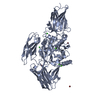 1nudC  1nufC  1l9nS S: Starting model for refinement C: citing same article ( |
|---|---|
| Similar structure data |
- Links
Links
- Assembly
| Deposited unit | 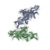
| ||||||||
|---|---|---|---|---|---|---|---|---|---|
| 1 | 
| ||||||||
| 2 | 
| ||||||||
| Unit cell |
|
- Components
| #1: Protein | Mass: 76670.500 Da / Num. of mol.: 2 / Mutation: F264L Source method: isolated from a genetically manipulated source Source: (gene. exp.)  Homo sapiens (human) / Gene: TGM3 / Plasmid: Bac-N_Blue, Invitrogen / Cell line (production host): SF9 / Production host: Homo sapiens (human) / Gene: TGM3 / Plasmid: Bac-N_Blue, Invitrogen / Cell line (production host): SF9 / Production host:  References: UniProt: Q08188, protein-glutamine gamma-glutamyltransferase #2: Chemical | ChemComp-CL / #3: Chemical | ChemComp-CA / #4: Chemical | #5: Water | ChemComp-HOH / | |
|---|
-Experimental details
-Experiment
| Experiment | Method:  X-RAY DIFFRACTION / Number of used crystals: 1 X-RAY DIFFRACTION / Number of used crystals: 1 |
|---|
- Sample preparation
| Crystal | Density Matthews: 2.86 Å3/Da / Density % sol: 57.05 % | ||||||||||||||||||||||||||||||||||||||||||||||||||||||||||||||||||||||||||||||||||||
|---|---|---|---|---|---|---|---|---|---|---|---|---|---|---|---|---|---|---|---|---|---|---|---|---|---|---|---|---|---|---|---|---|---|---|---|---|---|---|---|---|---|---|---|---|---|---|---|---|---|---|---|---|---|---|---|---|---|---|---|---|---|---|---|---|---|---|---|---|---|---|---|---|---|---|---|---|---|---|---|---|---|---|---|---|---|
| Crystal grow | Temperature: 288 K / Method: vapor diffusion, hanging drop / pH: 7.5 Details: 100 mM NaCl, 100 mM NaHEPES (pH 7.5) 4-8% Peg 4K, VAPOR DIFFUSION, HANGING DROP, temperature 288K | ||||||||||||||||||||||||||||||||||||||||||||||||||||||||||||||||||||||||||||||||||||
| Crystal grow | *PLUS Temperature: 21 ℃ / pH: 8 | ||||||||||||||||||||||||||||||||||||||||||||||||||||||||||||||||||||||||||||||||||||
| Components of the solutions | *PLUS
|
-Data collection
| Diffraction |
| ||||||||||||||||||
|---|---|---|---|---|---|---|---|---|---|---|---|---|---|---|---|---|---|---|---|
| Diffraction source |
| ||||||||||||||||||
| Detector |
| ||||||||||||||||||
| Radiation | Monochromator: SI 111 channel / Protocol: SINGLE WAVELENGTH / Monochromatic (M) / Laue (L): M / Scattering type: x-ray | ||||||||||||||||||
| Radiation wavelength |
| ||||||||||||||||||
| Reflection | Resolution: 2.4→20 Å / Num. obs: 65766 / % possible obs: 10.2 % / Observed criterion σ(F): 2 / Observed criterion σ(I): 2 / Biso Wilson estimate: 18.1 Å2 | ||||||||||||||||||
| Reflection shell | Resolution: 2.4→2.55 Å / % possible all: 97.7 | ||||||||||||||||||
| Reflection | *PLUS Lowest resolution: 25 Å / % possible obs: 98.3 % / Num. measured all: 495285 | ||||||||||||||||||
| Reflection shell | *PLUS % possible obs: 97.7 % / Mean I/σ(I) obs: 2 |
- Processing
| Software |
| |||||||||||||||||||||
|---|---|---|---|---|---|---|---|---|---|---|---|---|---|---|---|---|---|---|---|---|---|---|
| Refinement | Method to determine structure:  MOLECULAR REPLACEMENT MOLECULAR REPLACEMENTStarting model: PDB Entry 1L9N Resolution: 2.4→20 Å / Rfactor Rfree error: 0.003 / Isotropic thermal model: Isotropic / Cross valid method: THROUGHOUT / σ(F): 0 / Stereochemistry target values: Engh & Huber
| |||||||||||||||||||||
| Displacement parameters | Biso mean: 24.4 Å2
| |||||||||||||||||||||
| Refine analyze |
| |||||||||||||||||||||
| Refinement step | Cycle: LAST / Resolution: 2.4→20 Å
| |||||||||||||||||||||
| Refine LS restraints |
| |||||||||||||||||||||
| LS refinement shell | Resolution: 2.4→2.55 Å / Rfactor Rfree error: 0.011
| |||||||||||||||||||||
| Refinement | *PLUS Lowest resolution: 25 Å / % reflection Rfree: 10 % | |||||||||||||||||||||
| Solvent computation | *PLUS | |||||||||||||||||||||
| Displacement parameters | *PLUS | |||||||||||||||||||||
| Refine LS restraints | *PLUS
|
 Movie
Movie Controller







 PDBj
PDBj


