 Movie
Movie Controller

[English] 日本語
 Yorodumi
- PDB-1l9m: Three-dimensional structure of the human transglutaminase 3 enzym... -
+ Open data

- Basic information
| Entry | Database: PDB / ID: 1l9m | ||||||
|---|---|---|---|---|---|---|---|
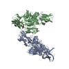
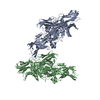
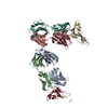
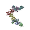
| Title | Three-dimensional structure of the human transglutaminase 3 enzyme: binding of calcium ions change structure for activation | ||||||
 Components Components | Protein-glutamine glutamyltransferase E3 | ||||||
 Keywords Keywords | TRANSFERASE / Activation / Calcium binding / transglutaminase | ||||||
| Function / homology |  Function and homology information Function and homology informationprotein-glutamine gamma-glutamyltransferase / protein-glutamine gamma-glutamyltransferase activity / peptide cross-linking / hair follicle morphogenesis / extrinsic component of cytoplasmic side of plasma membrane / acyltransferase activity / keratinization / catalytic activity / keratinocyte differentiation / protein modification process ...protein-glutamine gamma-glutamyltransferase / protein-glutamine gamma-glutamyltransferase activity / peptide cross-linking / hair follicle morphogenesis / extrinsic component of cytoplasmic side of plasma membrane / acyltransferase activity / keratinization / catalytic activity / keratinocyte differentiation / protein modification process / calcium ion binding / structural molecule activity / protein-containing complex / extracellular exosome / cytoplasm Similarity search - Function | ||||||
| Biological species |  Homo sapiens (human) Homo sapiens (human) | ||||||
| Method |  X-RAY DIFFRACTION / SYNCHROTRON / MOLECULAR REPLACEMENT / Resolution: 2.1 Å X-RAY DIFFRACTION / SYNCHROTRON / MOLECULAR REPLACEMENT / Resolution: 2.1 Å | ||||||
 Authors Authors | Ahvazi, B. | ||||||
 Citation Citation | Journal: EMBO J. / Year: 2002 Title: Three-dimensional structure of the human transglutaminase 3 enzyme: binding of calcium ions changes structure for activation. Authors: Ahvazi, B. / Kim, H.C. / Kee, S.H. / Nemes, Z. / Steinert, P.M. #1: Journal: FEBS Lett. / Year: 1998Title: Two non-proline cis peptide bonds may be important for factor XIII function Authors: Yee, V.C. / Pedersen, L.C. / Le Trong, I. / Bishop, P.D. / Stenkamp, R.E. / Teller, D.C. #2: Journal: Proc.Natl.Acad.Sci.USA / Year: 1994Title: Three-dimensional structure of a transglutaminase: human blood coagulation factor XII Authors: Weiss, M.S. / Metzner, H.J. / Hilgenfeld, R. | ||||||
| History |
| ||||||
| Remark 999 | The following residues are noted as conflicts in the Swiss-Prot database: K12T, K561R, G654R. ... The following residues are noted as conflicts in the Swiss-Prot database: K12T, K561R, G654R. According to the author, residue 250 is Asp and does not represent a mutation but a mistake in the Swiss-Prot database. |
- Structure visualization
| Structure viewer | Molecule: MolmilJmol/JSmol |
|---|
- Downloads & links
-Download
| PDBx/mmCIF format | 1l9m.cif.gz | 289.8 KB | Display | PDBx/mmCIF format |
|---|---|---|---|---|
| PDB format | pdb1l9m.ent.gz | 230.4 KB | Display | PDB format |
| PDBx/mmJSON format | 1l9m.json.gz | Tree view | PDBx/mmJSON format | |
| Others |  Other downloads Other downloads |
-Validation report
| Arichive directory | https://data.pdbj.org/pub/pdb/validation_reports/l9/1l9mftp://data.pdbj.org/pub/pdb/validation_reports/l9/1l9m | HTTPS FTP |
|---|
-Related structure data
| Related structure data |  1l9nC 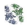 1ggtS C: citing same article ( S: Starting model for refinement |
|---|---|
| Similar structure data |
-Links
 PDBj
PDBj


- Assembly
| Deposited unit | 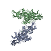
| ||||||||
|---|---|---|---|---|---|---|---|---|---|
| 1 |
| ||||||||
| Unit cell |
|
-Components
| #1: Protein | Mass: 76670.500 Da / Num. of mol.: 2 / Mutation: F264L Source method: isolated from a genetically manipulated source Source: (gene. exp.) Homo sapiens (human) / Tissue: Foreskin / Gene: TGM3 / Plasmid: Bac-N-Blue, Invitrogen / Cell line (production host): SF9 / Production host:   Spodoptera frugiperda (fall armyworm) / Strain (production host): baculovirus system Spodoptera frugiperda (fall armyworm) / Strain (production host): baculovirus systemReferences: UniProt: Q08188, protein-glutamine gamma-glutamyltransferase #2: Chemical |   Mass: 79.904 Da / Num. of mol.: 2 / Source method: obtained synthetically / Formula: Br Mass: 79.904 Da / Num. of mol.: 2 / Source method: obtained synthetically / Formula: Br#3: Chemical |   Mass: 35.453 Da / Num. of mol.: 2 / Source method: obtained synthetically / Formula: Cl Mass: 35.453 Da / Num. of mol.: 2 / Source method: obtained synthetically / Formula: Cl#4: Chemical |   Mass: 40.078 Da / Num. of mol.: 2 / Source method: obtained synthetically / Formula: Ca Mass: 40.078 Da / Num. of mol.: 2 / Source method: obtained synthetically / Formula: Ca#5: Water | ChemComp-HOH / |  Mass: 18.015 Da / Num. of mol.: 901 / Source method: isolated from a natural source / Formula: H2O Mass: 18.015 Da / Num. of mol.: 901 / Source method: isolated from a natural source / Formula: H2O |
|---|
-Experimental details
-Experiment
| Experiment | Method: X-RAY DIFFRACTION / Number of used crystals: 3 |
|---|
- Sample preparation
| Crystal | Density Matthews: 2.89 Å3/Da / Density % sol: 57.51 % | ||||||||||||||||||||||||||||||||||||||||||||||||||||||||
|---|---|---|---|---|---|---|---|---|---|---|---|---|---|---|---|---|---|---|---|---|---|---|---|---|---|---|---|---|---|---|---|---|---|---|---|---|---|---|---|---|---|---|---|---|---|---|---|---|---|---|---|---|---|---|---|---|---|
| Crystal grow | Temperature: 288 K / Method: vapor diffusion, hanging drop / pH: 8.5 Details: 4%(W/V) Peg 20K, 100 mM Tris_HCl(pH 8.5), VAPOR DIFFUSION, HANGING DROP, temperature 288K | ||||||||||||||||||||||||||||||||||||||||||||||||||||||||
| Crystal grow | *PLUS Temperature: 15 ℃ / pH: 8 | ||||||||||||||||||||||||||||||||||||||||||||||||||||||||
| Components of the solutions | *PLUS
|
-Data collection
| Diffraction |
| ||||||||||||||||||
|---|---|---|---|---|---|---|---|---|---|---|---|---|---|---|---|---|---|---|---|
| Diffraction source |
| ||||||||||||||||||
| Detector |
| ||||||||||||||||||
| Radiation | Monochromator: SI 111 channel / Protocol: SINGLE WAVELENGTH / Monochromatic (M) / Laue (L): M / Scattering type: x-ray | ||||||||||||||||||
| Radiation wavelength |
| ||||||||||||||||||
| Reflection | Resolution: 2.1→20 Å / Num. obs: 75013 / % possible obs: 93 % / Observed criterion σ(I): -3 / Biso Wilson estimate: 15.2 Å2 / Net I/σ(I): 11.3 | ||||||||||||||||||
| Reflection shell | Resolution: 2.1→20 Å / % possible all: 93 | ||||||||||||||||||
| Reflection | *PLUS Highest resolution: 2.2 Å / % possible obs: 94.6 % / Num. measured all: 521626 | ||||||||||||||||||
| Reflection shell | *PLUS Highest resolution: 2.2 Å / Lowest resolution: 2.28 Å / % possible obs: 93 % / Mean I/σ(I) obs: 2.9 |

- Processing
| Software |
| |||||||||||||||||||||||||||
|---|---|---|---|---|---|---|---|---|---|---|---|---|---|---|---|---|---|---|---|---|---|---|---|---|---|---|---|---|
| Refinement | Method to determine structure: MOLECULAR REPLACEMENT Starting model: PDB ENTRY 1GGT Resolution: 2.1→20 Å / Isotropic thermal model: Isotropic / Cross valid method: THROUGHOUT / σ(F): 0 / Stereochemistry target values: Engh & Huber Details: NCS two fold averaging was employed during refinement
| |||||||||||||||||||||||||||
| Displacement parameters | Biso mean: 26.3 Å2
| |||||||||||||||||||||||||||
| Refine analyze |
| |||||||||||||||||||||||||||
| Refinement step | Cycle: LAST / Resolution: 2.1→20 Å
| |||||||||||||||||||||||||||
| Refine LS restraints |
| |||||||||||||||||||||||||||
| LS refinement shell | Resolution: 2.1→2.34 Å / Rfactor Rfree error: 0.009
| |||||||||||||||||||||||||||
| Refinement | *PLUS Highest resolution: 2.2 Å / Lowest resolution: 20 Å / % reflection Rfree: 10 % / Rfactor obs: 0.182 / Rfactor Rfree: 0.225 / Rfactor Rwork: 0.182 | |||||||||||||||||||||||||||
| Solvent computation | *PLUS | |||||||||||||||||||||||||||
| Displacement parameters | *PLUS | |||||||||||||||||||||||||||
| Refine LS restraints | *PLUS
| |||||||||||||||||||||||||||
| LS refinement shell | *PLUS Rfactor obs: 0.248 |
