 Movie
Movie Controller
 Structure viewers
 About Yorodumi Papers


+Search query
-Structure paper
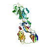
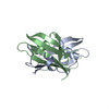
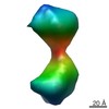
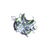
| Title | Assembly, maturation and three-dimensional helical structure of the teratogenic rubella virus. |
|---|---|
| Journal, issue, pages | PLoS Pathog, Vol. 13, Issue 6, Page e1006377, Year 2017 |
| Publish date | Jun 2, 2017 |
 Authors Authors | Vidya Mangala Prasad / Thomas Klose / Michael G Rossmann /  |
| PubMed Abstract | Viral infections during pregnancy are a significant cause of infant morbidity and mortality. Of these, rubella virus infection is a well-substantiated example that leads to miscarriages or severe ...Viral infections during pregnancy are a significant cause of infant morbidity and mortality. Of these, rubella virus infection is a well-substantiated example that leads to miscarriages or severe fetal defects. However, structural information about the rubella virus has been lacking due to the pleomorphic nature of the virions. Here we report a helical structure of rubella virions using cryo-electron tomography. Sub-tomogram averaging of the surface spikes established the relative positions of the viral glycoproteins, which differed from the earlier icosahedral models of the virus. Tomographic analyses of in vitro assembled nucleocapsids and virions provide a template for viral assembly. Comparisons of immature and mature virions show large rearrangements in the glycoproteins that may be essential for forming the infectious virions. These results present the first known example of a helical membrane-enveloped virus, while also providing a structural basis for its assembly and maturation pathway. |
 External links External links | PLoS Pathog / PubMed:28575072 / PubMed Central |
| Methods | EM (subtomogram averaging) |
| Resolution | 11.1 - 35.0 Å |
| Structure data | EMDB-8248: Subtomogram-averaged surface glycoprotein spike density of rubella virus EMDB-8249: Subtomogram-averaged map of a single rubella virus capsid unit EMDB-8250: Subtomogram-averaged map of a single rubella virus capsid unit  EMDB-8251: |
| Chemicals |  ChemComp-HOH: |
| Source |
|
 Keywords Keywords | VIRAL PROTEIN / Rubella virus / surface glycoprotein spike / E1-E2 heterodimer / rubella virus capsid protein |


 rubella virus