- EMDB-4692: Human-D02 Nucleosome Core Particle with biotin-streptavidin label -
+
データを開く
IDまたはキーワード:
読み込み中...
-
基本情報
登録情報
データベース: EMDB / ID: EMD-4692
タイトル
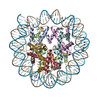
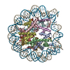
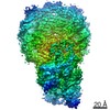
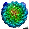
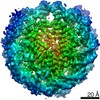
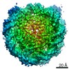
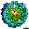
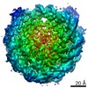
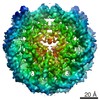
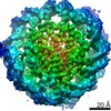
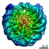
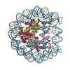
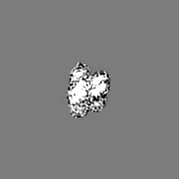
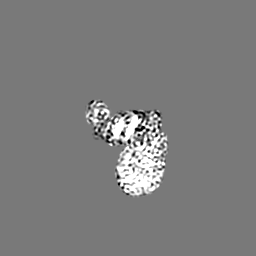
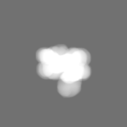
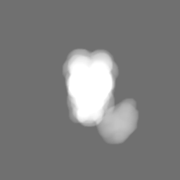
Human-D02 Nucleosome Core Particle with biotin-streptavidin label
マップデータ
human nucleosome core particle wrapped with 145bp of D02 DNA with biotin-streptavidin at distal end
試料
複合体: Human-D02 Nucleosome Core Particle with biotin-streptavidin label
複合体: Histones
タンパク質・ペプチド: Histone H3.3
タンパク質・ペプチド: Histone H4
タンパク質・ペプチド: Histone H2A type 1
タンパク質・ペプチド: Histone H2B type 1-C/E/F/G/I
複合体: DNA
DNA: DNA (142-MER)
DNA: DNA (142-MER)
リガンド: MANGANESE (II) ION
キーワード
chromatin / nucleosome / retrovirus / DNA BINDING PROTEIN
機能・相同性
機能・相同性情報
Barr body / negative regulation of chromosome condensation / inner kinetochore / pericentric heterochromatin formation / muscle cell differentiation / oocyte maturation / nucleosomal DNA binding / nucleus organization / spermatid development / single fertilization ...Barr body / negative regulation of chromosome condensation / inner kinetochore / pericentric heterochromatin formation / muscle cell differentiation / oocyte maturation / nucleosomal DNA binding / nucleus organization / spermatid development / single fertilization / subtelomeric heterochromatin formation / RNA polymerase II core promoter sequence-specific DNA binding / negative regulation of megakaryocyte differentiation / protein localization to CENP-A containing chromatin / Replacement of protamines by nucleosomes in the male pronucleus / CENP-A containing nucleosome / Packaging Of Telomere Ends / Recognition and association of DNA glycosylase with site containing an affected purine / Cleavage of the damaged purine / embryo implantation / Deposition of new CENPA-containing nucleosomes at the centromere / telomere organization / Recognition and association of DNA glycosylase with site containing an affected pyrimidine / Cleavage of the damaged pyrimidine / RNA Polymerase I Promoter Opening / Inhibition of DNA recombination at telomere / Assembly of the ORC complex at the origin of replication / Meiotic synapsis / SUMOylation of chromatin organization proteins / Regulation of endogenous retroelements by the Human Silencing Hub (HUSH) complex / DNA methylation / Condensation of Prophase Chromosomes / Chromatin modifications during the maternal to zygotic transition (MZT) / SIRT1 negatively regulates rRNA expression / HCMV Late Events / ERCC6 (CSB) and EHMT2 (G9a) positively regulate rRNA expression / PRC2 methylates histones and DNA / innate immune response in mucosa / Regulation of endogenous retroelements by KRAB-ZFP proteins / Defective pyroptosis / HDACs deacetylate histones / Regulation of endogenous retroelements by Piwi-interacting RNAs (piRNAs) / Nonhomologous End-Joining (NHEJ) / RNA Polymerase I Promoter Escape / Transcriptional regulation by small RNAs / Formation of the beta-catenin:TCF transactivating complex / Activated PKN1 stimulates transcription of AR (androgen receptor) regulated genes KLK2 and KLK3 / HDMs demethylate histones / RUNX1 regulates genes involved in megakaryocyte differentiation and platelet function / G2/M DNA damage checkpoint / Negative Regulation of CDH1 Gene Transcription / NoRC negatively regulates rRNA expression / PKMTs methylate histone lysines / B-WICH complex positively regulates rRNA expression / DNA Damage/Telomere Stress Induced Senescence / Pre-NOTCH Transcription and Translation / male gonad development / Meiotic recombination / Activation of anterior HOX genes in hindbrain development during early embryogenesis / multicellular organism growth / Transcriptional regulation of granulopoiesis / Metalloprotease DUBs / RMTs methylate histone arginines / HCMV Early Events / osteoblast differentiation / structural constituent of chromatin / UCH proteinases / nucleosome / antimicrobial humoral immune response mediated by antimicrobial peptide / heterochromatin formation / nucleosome assembly / antibacterial humoral response / E3 ubiquitin ligases ubiquitinate target proteins / HATs acetylate histones / Recruitment and ATM-mediated phosphorylation of repair and signaling proteins at DNA double strand breaks / Factors involved in megakaryocyte development and platelet production / MLL4 and MLL3 complexes regulate expression of PPARG target genes in adipogenesis and hepatic steatosis / chromatin organization / RUNX1 regulates transcription of genes involved in differentiation of HSCs / positive regulation of cell growth / Processing of DNA double-strand break ends / Senescence-Associated Secretory Phenotype (SASP) / Oxidative Stress Induced Senescence / Estrogen-dependent gene expression / chromosome, telomeric region / cell population proliferation / Ub-specific processing proteases / defense response to Gram-positive bacterium / RNA polymerase II cis-regulatory region sequence-specific DNA binding / Amyloid fiber formation / protein heterodimerization activity / enzyme binding / protein-containing complex / : / DNA binding / RNA binding / extracellular exosome / extracellular region / nucleoplasm / membrane 類似検索 - 分子機能
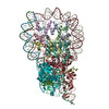
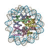
ジャーナル: Nat Commun / 年: 2019 タイトル: Retroviral integration into nucleosomes through DNA looping and sliding along the histone octamer. 著者: Marcus D Wilson / Ludovic Renault / Daniel P Maskell / Mohamed Ghoneim / Valerie E Pye / Andrea Nans / David S Rueda / Peter Cherepanov / Alessandro Costa / 要旨: Retroviral integrase can efficiently utilise nucleosomes for insertion of the reverse-transcribed viral DNA. In face of the structural constraints imposed by the nucleosomal structure, integrase ...Retroviral integrase can efficiently utilise nucleosomes for insertion of the reverse-transcribed viral DNA. In face of the structural constraints imposed by the nucleosomal structure, integrase gains access to the scissile phosphodiester bonds by lifting DNA off the histone octamer at the site of integration. To clarify the mechanism of DNA looping by integrase, we determined a 3.9 Å resolution structure of the prototype foamy virus intasome engaged with a nucleosome core particle. The structural data along with complementary single-molecule Förster resonance energy transfer measurements reveal twisting and sliding of the nucleosomal DNA arm proximal to the integration site. Sliding the nucleosomal DNA by approximately two base pairs along the histone octamer accommodates the necessary DNA lifting from the histone H2A-H2B subunits to allow engagement with the intasome. Thus, retroviral integration into nucleosomes involves the looping-and-sliding mechanism for nucleosomal DNA repositioning, bearing unexpected similarities to chromatin remodelers.
全体 : Human-D02 Nucleosome Core Particle with biotin-streptavidin label
全体
名称: Human-D02 Nucleosome Core Particle with biotin-streptavidin label
要素
複合体: Human-D02 Nucleosome Core Particle with biotin-streptavidin label
複合体: Histones
タンパク質・ペプチド: Histone H3.3
タンパク質・ペプチド: Histone H4
タンパク質・ペプチド: Histone H2A type 1
タンパク質・ペプチド: Histone H2B type 1-C/E/F/G/I
複合体: DNA
DNA: DNA (142-MER)
DNA: DNA (142-MER)
リガンド: MANGANESE (II) ION
+
超分子 #1: Human-D02 Nucleosome Core Particle with biotin-streptavidin label
超分子
名称: Human-D02 Nucleosome Core Particle with biotin-streptavidin label タイプ: complex / ID: 1 / 親要素: 0 / 含まれる分子: #1-#6 詳細: human histones refolded as an octamer with native human D02 sequence with flexible linker biotin.tetravalent streptavidin added onto refolded nucleosomes and sample crosslinked with glutaraldehyde
タイプ: MAXIMUM LIKELIHOOD / ソフトウェア - 名称: RELION (ver. 2.1)
最終 角度割当
タイプ: MAXIMUM LIKELIHOOD / ソフトウェア - 名称: RELION (ver. 2.1)
最終 3次元分類
クラス数: 2 / 平均メンバー数/クラス: 64000 / ソフトウェア - 名称: RELION (ver. 2.1) 詳細: Two rounds of 3D classification. First with 8 classes, second with 2 classes. Slight conformational difference between two classes.
Chain - Source name: PDB / Chain - Initial model type: experimental model
詳細
The initial model was placed in the density using Chimera. Manual building was performed in Coot and final refinement was carried out using phenix.real_space_refine. Additional restraints describing protein secondary structure, DNA base pairing and stacking were used in Phenix.
精密化
空間: REAL
得られたモデル
PDB-6r0c: Human-D02 Nucleosome Core Particle with biotin-streptavidin label
 ムービー
ムービー コントローラー


 データを開く
 基本情報
基本情報 マップデータ
 試料
試料 キーワード
キーワード 機能・相同性情報
 Homo sapiens (ヒト)
 データ登録者
 英国, 2件
  引用
引用
 構造の表示
構造の表示
 ダウンロードとリンク
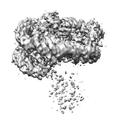 emd_4692.png
 http://ftp.pdbj.org/pub/emdb/structures/EMD-4692
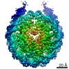


 Z (Sec.)
 Y (Row.)
 X (Col.)


 試料の構成要素
 解析
解析 電子顕微鏡法
 FIELD EMISSION GUN