National Institutes of Health/National Institute of General Medical Sciences (NIH/NIGMS)
P41GM103832
米国
National Institutes of Health/National Institute of General Medical Sciences (NIH/NIGMS)
R01GM079429
米国
National Institutes of Health/National Institute Of Allergy and Infectious Diseases (NIH/NIAID)
P01AI120943
米国
National Institutes of Health/National Institute of General Medical Sciences (NIH/NIGMS)
S10OD021600
米国
引用
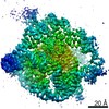
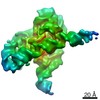
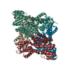
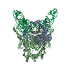
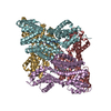
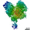
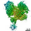
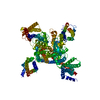
ジャーナル: Nature / 年: 2021 タイトル: Cryo-EM structures of full-length Tetrahymena ribozyme at 3.1 Å resolution. 著者: Zhaoming Su / Kaiming Zhang / Kalli Kappel / Shanshan Li / Michael Z Palo / Grigore D Pintilie / Ramya Rangan / Bingnan Luo / Yuquan Wei / Rhiju Das / Wah Chiu / 要旨: Single-particle cryogenic electron microscopy (cryo-EM) has become a standard technique for determining protein structures at atomic resolution. However, cryo-EM studies of protein-free RNA are in ...Single-particle cryogenic electron microscopy (cryo-EM) has become a standard technique for determining protein structures at atomic resolution. However, cryo-EM studies of protein-free RNA are in their early days. The Tetrahymena thermophila group I self-splicing intron was the first ribozyme to be discovered and has been a prominent model system for the study of RNA catalysis and structure-function relationships, but its full structure remains unknown. Here we report cryo-EM structures of the full-length Tetrahymena ribozyme in substrate-free and bound states at a resolution of 3.1 Å. Newly resolved peripheral regions form two coaxially stacked helices; these are interconnected by two kissing loop pseudoknots that wrap around the catalytic core and include two previously unforeseen (to our knowledge) tertiary interactions. The global architecture is nearly identical in both states; only the internal guide sequence and guanosine binding site undergo a large conformational change and a localized shift, respectively, upon binding of RNA substrates. These results provide a long-sought structural view of a paradigmatic RNA enzyme and signal a new era for the cryo-EM-based study of structure-function relationships in ribozymes.
 ムービー
ムービー コントローラー


 データを開く
 基本情報
基本情報 マップデータ
 試料
試料 キーワード
キーワード
 Tetrahymena thermophila (真核生物)
 データ登録者
 中国,
中国,  米国, 6件
  引用
引用 構造の表示
構造の表示 ムービービューア
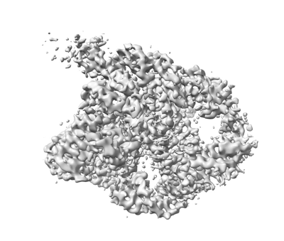
 ダウンロードとリンク
 emd_31385.png
 http://ftp.pdbj.org/pub/emdb/structures/EMD-31385
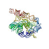


 Z (Sec.)
 Y (Row.)
 X (Col.)


 試料の構成要素
 解析
解析 電子顕微鏡法
 FIELD EMISSION GUN

