 Movie
Movie Controller


+ Open data

- Basic information
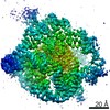
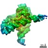
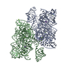
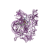
| Entry | Database: PDB / ID: 7ez0 | |||||||||||||||||||||
|---|---|---|---|---|---|---|---|---|---|---|---|---|---|---|---|---|---|---|---|---|---|---|
| Title | Apo L-21 ScaI Tetrahymena ribozyme | |||||||||||||||||||||
 Components Components | Apo L-21 ScaI Tetrahymena ribozyme | |||||||||||||||||||||
 Keywords Keywords | RNA / RNA structure / Tetrahymena ribozyme | |||||||||||||||||||||
| Function / homology | : / RNA / RNA (> 10) / RNA (> 100) Function and homology information Function and homology information | |||||||||||||||||||||
| Biological species |   Tetrahymena thermophila (eukaryote) Tetrahymena thermophila (eukaryote) | |||||||||||||||||||||
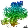
| Method | ELECTRON MICROSCOPY / single particle reconstruction / cryo EM / Resolution: 3.14 Å | |||||||||||||||||||||
 Authors Authors | Su, Z. / Zhang, K. / Kappel, K. / Luo, B. / Das, R. / Chiu, W. | |||||||||||||||||||||
| Funding support |  China, China,  United States, 6items United States, 6items
| |||||||||||||||||||||
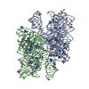
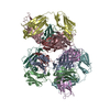
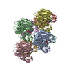
 Citation Citation | Journal: Nature / Year: 2021 Title: Cryo-EM structures of full-length Tetrahymena ribozyme at 3.1 Å resolution. Authors: Zhaoming Su / Kaiming Zhang / Kalli Kappel / Shanshan Li / Michael Z Palo / Grigore D Pintilie / Ramya Rangan / Bingnan Luo / Yuquan Wei / Rhiju Das / Wah Chiu / Abstract: Single-particle cryogenic electron microscopy (cryo-EM) has become a standard technique for determining protein structures at atomic resolution. However, cryo-EM studies of protein-free RNA are in ...Single-particle cryogenic electron microscopy (cryo-EM) has become a standard technique for determining protein structures at atomic resolution. However, cryo-EM studies of protein-free RNA are in their early days. The Tetrahymena thermophila group I self-splicing intron was the first ribozyme to be discovered and has been a prominent model system for the study of RNA catalysis and structure-function relationships, but its full structure remains unknown. Here we report cryo-EM structures of the full-length Tetrahymena ribozyme in substrate-free and bound states at a resolution of 3.1 Å. Newly resolved peripheral regions form two coaxially stacked helices; these are interconnected by two kissing loop pseudoknots that wrap around the catalytic core and include two previously unforeseen (to our knowledge) tertiary interactions. The global architecture is nearly identical in both states; only the internal guide sequence and guanosine binding site undergo a large conformational change and a localized shift, respectively, upon binding of RNA substrates. These results provide a long-sought structural view of a paradigmatic RNA enzyme and signal a new era for the cryo-EM-based study of structure-function relationships in ribozymes. | |||||||||||||||||||||
| History |
|
- Structure visualization
| Movie |
Movie viewer |
|---|---|
| Structure viewer | Molecule: MolmilJmol/JSmol |
- Downloads & links
-Download
| PDBx/mmCIF format | 7ez0.cif.gz | 193.7 KB | Display | PDBx/mmCIF format |
|---|---|---|---|---|
| PDB format | pdb7ez0.ent.gz | 143.1 KB | Display | PDB format |
| PDBx/mmJSON format | 7ez0.json.gz | Tree view | PDBx/mmJSON format | |
| Others |  Other downloads Other downloads |
-Validation report
| Arichive directory | https://data.pdbj.org/pub/pdb/validation_reports/ez/7ez0ftp://data.pdbj.org/pub/pdb/validation_reports/ez/7ez0 | HTTPS FTP |
|---|
-Related structure data
| Related structure data |  31385MC  7ez2C M: map data used to model this data C: citing same article ( |
|---|---|
| Similar structure data |


-Links
 PDBj
PDBj


- Assembly
| Deposited unit | 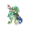
|
|---|---|
| 1 |
|
-Components
| #1: RNA chain | Mass: 125096.773 Da / Num. of mol.: 1 Source method: isolated from a genetically manipulated source Source: (gene. exp.) Tetrahymena thermophila (eukaryote) / Production host: synthetic construct (others) / References: GenBank: 10832 | ||
|---|---|---|---|
| #2: Chemical | ChemComp-MG /   Mass: 24.305 Da / Num. of mol.: 27 / Source method: obtained synthetically / Formula: Mg Mass: 24.305 Da / Num. of mol.: 27 / Source method: obtained synthetically / Formula: MgHas ligand of interest | N | |
-Experimental details
-Experiment
| Experiment | Method: ELECTRON MICROSCOPY |
|---|---|
| EM experiment | Aggregation state: PARTICLE / 3D reconstruction method: single particle reconstruction |
- Sample preparation
| Component | Name: Apo L-21 ScaI Tetrahymena ribozyme with metal ions. / Type: COMPLEX / Entity ID: #1 / Source: RECOMBINANT |
|---|---|
| Molecular weight | Experimental value: NO |
| Source (natural) | Organism: Tetrahymena thermophila (eukaryote) |
| Source (recombinant) | Organism: synthetic construct (others) |
| Buffer solution | pH: 8.1 |
| Specimen | Conc.: 3 mg/ml / Embedding applied: NO / Shadowing applied: NO / Staining applied: NO / Vitrification applied: YES |
| Specimen support | Grid material: COPPER / Grid mesh size: 200 divisions/in. / Grid type: Quantifoil R2/1 |
| Vitrification | Instrument: FEI VITROBOT MARK IV / Cryogen name: ETHANE / Humidity: 100 % / Chamber temperature: 278 K |
- Electron microscopy imaging
| Experimental equipment |  Model: Titan Krios / Image courtesy: FEI Company |
|---|---|
| Microscopy | Model: FEI TITAN KRIOS |
| Electron gun | Electron source:  FIELD EMISSION GUN / Accelerating voltage: 300 kV / Illumination mode: FLOOD BEAM FIELD EMISSION GUN / Accelerating voltage: 300 kV / Illumination mode: FLOOD BEAM |
| Electron lens | Mode: BRIGHT FIELD / Nominal magnification: 215000 X / Nominal defocus max: 1500 nm / Nominal defocus min: 300 nm / Cs: 2.7 mm / C2 aperture diameter: 50 µm / Alignment procedure: COMA FREE |
| Specimen holder | Cryogen: NITROGEN / Specimen holder model: FEI TITAN KRIOS AUTOGRID HOLDER / Temperature (min): 78.2 K |
| Image recording | Electron dose: 75 e/Å2 / Detector mode: COUNTING / Film or detector model: GATAN K2 QUANTUM (4k x 4k) / Num. of real images: 7577 |
| EM imaging optics | Energyfilter name: GIF Bioquantum / Energyfilter slit width: 20 eV |
| Image scans | Sampling size: 5 µm / Width: 3838 / Height: 3710 / Movie frames/image: 25 / Used frames/image: 1-25 |
- Processing
| EM software |
| ||||||||||||||||||||||||||||||||||||||||||||
|---|---|---|---|---|---|---|---|---|---|---|---|---|---|---|---|---|---|---|---|---|---|---|---|---|---|---|---|---|---|---|---|---|---|---|---|---|---|---|---|---|---|---|---|---|---|
| CTF correction | Type: PHASE FLIPPING AND AMPLITUDE CORRECTION | ||||||||||||||||||||||||||||||||||||||||||||
| Particle selection | Num. of particles selected: 1658961 | ||||||||||||||||||||||||||||||||||||||||||||
| Symmetry | Point symmetry: C1 (asymmetric) | ||||||||||||||||||||||||||||||||||||||||||||
| 3D reconstruction | Resolution: 3.14 Å / Resolution method: FSC 0.143 CUT-OFF / Num. of particles: 415918 / Algorithm: FOURIER SPACE / Num. of class averages: 1 / Symmetry type: POINT | ||||||||||||||||||||||||||||||||||||||||||||
| Atomic model building | Protocol: AB INITIO MODEL / Space: REAL | ||||||||||||||||||||||||||||||||||||||||||||
| Atomic model building | PDB-ID: 6WLS Pdb chain-ID: A / Accession code: 6WLS / Pdb chain residue range: 22-409 / Source name: PDB / Type: experimental model |

