[English] 日本語
 Yorodumi
- EMDB-2701: Human dynamin 1 K44A superconstricted polymer stabilized with GTP -
+ Open data
- Basic information
| Entry | Database: EMDB / ID: EMD-2701 | |||||||||
|---|---|---|---|---|---|---|---|---|---|---|
| Title | Human dynamin 1 K44A superconstricted polymer stabilized with GTP | |||||||||
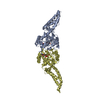
 Map data Map data | Reconstruction of a dynamin mutant, K44A, bound to DOPS lipid tube | |||||||||
 Sample Sample |
| |||||||||
 Keywords Keywords | Dynamin / endocytosis / membrane fission / GTPase / intracellular trafficking | |||||||||
| Function / homology |  Function and homology information Function and homology informationclathrin coat assembly involved in endocytosis / vesicle scission / synaptic vesicle budding from presynaptic endocytic zone membrane / dynamin GTPase / chromaffin granule / regulation of vesicle size / Retrograde neurotrophin signalling / Toll Like Receptor 4 (TLR4) Cascade / Formation of annular gap junctions / Gap junction degradation ...clathrin coat assembly involved in endocytosis / vesicle scission / synaptic vesicle budding from presynaptic endocytic zone membrane / dynamin GTPase / chromaffin granule / regulation of vesicle size / Retrograde neurotrophin signalling / Toll Like Receptor 4 (TLR4) Cascade / Formation of annular gap junctions / Gap junction degradation / endosome organization / phosphatidylinositol-3,4,5-trisphosphate binding / Recycling pathway of L1 / EPH-ephrin mediated repulsion of cells / endocytic vesicle / phosphatidylinositol-4,5-bisphosphate binding / clathrin-coated pit / MHC class II antigen presentation / receptor-mediated endocytosis / cell projection / protein homooligomerization / receptor internalization / endocytosis / GDP binding / presynapse / Clathrin-mediated endocytosis / protein homotetramerization / microtubule binding / microtubule / GTPase activity / synapse / protein kinase binding / GTP binding / protein homodimerization activity / RNA binding / extracellular exosome / identical protein binding / plasma membrane / cytoplasm Similarity search - Function | |||||||||
| Biological species |  Homo sapiens (human) Homo sapiens (human) | |||||||||
| Method | helical reconstruction / cryo EM / Resolution: 12.5 Å | |||||||||
 Authors Authors | Sundborger AC / Fang S / Heymann JA / Ray P / Chappie JS / Hinshaw JE | |||||||||
 Citation Citation |  Journal: Cell Rep / Year: 2014 Journal: Cell Rep / Year: 2014Title: A dynamin mutant defines a superconstricted prefission state. Authors: Anna C Sundborger / Shunming Fang / Jürgen A Heymann / Pampa Ray / Joshua S Chappie / Jenny E Hinshaw /  Abstract: Dynamin is a 100 kDa GTPase that organizes into helical assemblies at the base of nascent clathrin-coated vesicles. Formation of these oligomers stimulates the intrinsic GTPase activity of dynamin, ...Dynamin is a 100 kDa GTPase that organizes into helical assemblies at the base of nascent clathrin-coated vesicles. Formation of these oligomers stimulates the intrinsic GTPase activity of dynamin, which is necessary for efficient membrane fission during endocytosis. Recent evidence suggests that the transition state of dynamin's GTP hydrolysis reaction serves as a key determinant of productive fission. Here, we present the structure of a transition-state-defective dynamin mutant K44A trapped in a prefission state at 12.5 Å resolution. This structure constricts to 3.7 nm, reaching the theoretical limit required for spontaneous membrane fission. Computational docking indicates that the ground-state conformation of the dynamin polymer is sufficient to achieve this superconstricted prefission state and reveals how a two-start helical symmetry promotes the most efficient packing of dynamin tetramers around the membrane neck. These data suggest a model for the assembly and regulation of the minimal dynamin fission machine. | |||||||||
| History |
|
- Structure visualization
| Movie |
 Movie viewer Movie viewer |
|---|---|
| Structure viewer | EM map:  SurfView SurfView Molmil Molmil Jmol/JSmol Jmol/JSmol |
| Supplemental images |
- Downloads & links
-EMDB archive
| Map data |  emd_2701.map.gz emd_2701.map.gz | 6.5 MB |  EMDB map data format EMDB map data format | |
|---|---|---|---|---|
| Header (meta data) |  emd-2701-v30.xml emd-2701-v30.xml emd-2701.xml emd-2701.xml | 13.4 KB 13.4 KB | Display Display |  EMDB header EMDB header |
| Images |  EMD-2701.png EMD-2701.png | 194.1 KB | ||
| Archive directory |  http://ftp.pdbj.org/pub/emdb/structures/EMD-2701 http://ftp.pdbj.org/pub/emdb/structures/EMD-2701 ftp://ftp.pdbj.org/pub/emdb/structures/EMD-2701 ftp://ftp.pdbj.org/pub/emdb/structures/EMD-2701 | HTTPS FTP |
-Validation report
| Summary document |  emd_2701_validation.pdf.gz emd_2701_validation.pdf.gz | 294.2 KB | Display |  EMDB validaton report EMDB validaton report |
|---|---|---|---|---|
| Full document |  emd_2701_full_validation.pdf.gz emd_2701_full_validation.pdf.gz | 293.3 KB | Display | |
| Data in XML |  emd_2701_validation.xml.gz emd_2701_validation.xml.gz | 6.1 KB | Display | |
| Arichive directory |  https://ftp.pdbj.org/pub/emdb/validation_reports/EMD-2701 https://ftp.pdbj.org/pub/emdb/validation_reports/EMD-2701 ftp://ftp.pdbj.org/pub/emdb/validation_reports/EMD-2701 ftp://ftp.pdbj.org/pub/emdb/validation_reports/EMD-2701 | HTTPS FTP |
-Related structure data
| Related structure data |  4uudMC  4uukMC M: atomic model generated by this map C: citing same article ( |
|---|---|
| Similar structure data |
- Links
Links
| EMDB pages |  EMDB (EBI/PDBe) / EMDB (EBI/PDBe) /  EMDataResource EMDataResource |
|---|---|
| Related items in Molecule of the Month |
- Map
Map
| File |  Download / File: emd_2701.map.gz / Format: CCP4 / Size: 29.8 MB / Type: IMAGE STORED AS FLOATING POINT NUMBER (4 BYTES) Download / File: emd_2701.map.gz / Format: CCP4 / Size: 29.8 MB / Type: IMAGE STORED AS FLOATING POINT NUMBER (4 BYTES) | ||||||||||||||||||||||||||||||||||||||||||||||||||||||||||||||||||||
|---|---|---|---|---|---|---|---|---|---|---|---|---|---|---|---|---|---|---|---|---|---|---|---|---|---|---|---|---|---|---|---|---|---|---|---|---|---|---|---|---|---|---|---|---|---|---|---|---|---|---|---|---|---|---|---|---|---|---|---|---|---|---|---|---|---|---|---|---|---|
| Annotation | Reconstruction of a dynamin mutant, K44A, bound to DOPS lipid tube | ||||||||||||||||||||||||||||||||||||||||||||||||||||||||||||||||||||
| Projections & slices | Image control
Images are generated by Spider. | ||||||||||||||||||||||||||||||||||||||||||||||||||||||||||||||||||||
| Voxel size | X=Y=Z: 2.55 Å | ||||||||||||||||||||||||||||||||||||||||||||||||||||||||||||||||||||
| Density |
| ||||||||||||||||||||||||||||||||||||||||||||||||||||||||||||||||||||
| Symmetry | Space group: 1 | ||||||||||||||||||||||||||||||||||||||||||||||||||||||||||||||||||||
| Details | EMDB XML:
CCP4 map header:
| ||||||||||||||||||||||||||||||||||||||||||||||||||||||||||||||||||||
-Supplemental data
- Sample components
-Entire : GTP-stablized human dynamin 1 K44A dynamin polymer bound to DOPS ...
| Entire | Name: GTP-stablized human dynamin 1 K44A dynamin polymer bound to DOPS lipid bilayer |
|---|---|
| Components |
|
-Supramolecule #1000: GTP-stablized human dynamin 1 K44A dynamin polymer bound to DOPS ...
| Supramolecule | Name: GTP-stablized human dynamin 1 K44A dynamin polymer bound to DOPS lipid bilayer type: sample / ID: 1000 / Oligomeric state: helical assembly of dynamin / Number unique components: 1 |
|---|
-Macromolecule #1: Human dynamin 1
| Macromolecule | Name: Human dynamin 1 / type: protein_or_peptide / ID: 1 / Oligomeric state: helical assembly / Recombinant expression: Yes |
|---|---|
| Source (natural) | Organism:  Homo sapiens (human) / synonym: Human Homo sapiens (human) / synonym: Human |
| Molecular weight | Experimental: 98 MDa / Theoretical: 97.3 MDa |
| Recombinant expression | Organism:  Recombinant plasmid: pBlueBacIII baculovirus expression vector |
| Sequence | UniProtKB: Dynamin-1 |
-Experimental details
-Structure determination
| Method | cryo EM |
|---|---|
 Processing Processing | helical reconstruction |
| Aggregation state | helical array |
- Sample preparation
| Concentration | 0.2 mg/mL |
|---|---|
| Buffer | pH: 7.2 Details: 20 mM Hepes, 150 mM NaCl, 1 mM EGTA, 1 mM DTT, 1 mM MgCl |
| Grid | Details: Quantifoil R3.5/1 200 mesh Cu Holey Carbon |
| Vitrification | Cryogen name: ETHANE / Chamber humidity: 90 % / Chamber temperature: 93 K / Instrument: LEICA EM GP / Method: 40 seconds pre-blot, 2 second blot before plunging |
- Electron microscopy
| Microscope | FEI POLARA 300 |
|---|---|
| Temperature | Min: 93 K / Max: 95 K / Average: 94 K |
| Alignment procedure | Legacy - Astigmatism: Objective lens astigmatism was corrected at 100,000 times magnification |
| Date | Dec 17, 2012 |
| Image recording | Category: FILM / Film or detector model: KODAK SO-163 FILM / Digitization - Scanner: NIKON SUPER COOLSCAN 9000 / Digitization - Sampling interval: 12.5 µm / Number real images: 357 / Average electron dose: 10 e/Å2 / Od range: 1.5 |
| Tilt angle min | 0 |
| Tilt angle max | 0 |
| Electron beam | Acceleration voltage: 200 kV / Electron source:  FIELD EMISSION GUN FIELD EMISSION GUN |
| Electron optics | Calibrated magnification: 49000 / Illumination mode: FLOOD BEAM / Imaging mode: BRIGHT FIELD / Cs: 2.26 mm / Nominal defocus max: 2.5 µm / Nominal defocus min: 0.5 µm / Nominal magnification: 49000 |
| Sample stage | Specimen holder: Liquid Nitrogen cooled / Specimen holder model: OTHER |
| Experimental equipment |  Model: Tecnai Polara / Image courtesy: FEI Company |
- Image processing
| Details | The particles were aligned using IHRSR |
|---|---|
| Final reconstruction | Applied symmetry - Helical parameters - Δz: 17.19 Å Applied symmetry - Helical parameters - Δ&Phi: 30.59 ° Applied symmetry - Helical parameters - Axial symmetry: C1 (asymmetric) Algorithm: OTHER / Resolution.type: BY AUTHOR / Resolution: 12.5 Å / Resolution method: OTHER / Software - Name: Spider Details: A total of 7525 helical segments were incorporated by the IHRSR algorithm into the final reconstruction after 50 cycles |
| CTF correction | Details: each image using bsoft |
-Atomic model buiding 1
| Initial model | PDB ID: |
|---|---|
| Software | Name: YUP |
| Details | Initial fitting was performed using GG-GMPPCP monomers (PDB: 3ZYC), dynamin middle/GED stalk monomers excised from (PDB: 3SNH), and PH domain monomers (PDB: 1DYN). All-atom structures were refined using the YUP.SCX method of the YUP software package. |
| Refinement | Space: REAL / Protocol: FLEXIBLE FIT |
| Output model |  PDB-4uud:  PDB-4uuk: |
-Atomic model buiding 2
| Initial model | PDB ID: |
|---|---|
| Software | Name: YUP |
| Details | Initial fitting was performed using GG-GMPPCP monomers (PDB: 3ZYC), dynamin middle/GED stalk monomers excised from (PDB: 3SNH), and PH domain monomers (PDB: 1DYN). All-atom structures were refined using the YUP.SCX method of the YUP software package. |
| Refinement | Space: REAL / Protocol: FLEXIBLE FIT |
| Output model |  PDB-4uud:  PDB-4uuk: |
-Atomic model buiding 3
| Initial model | PDB ID: |
|---|---|
| Software | Name: YUP |
| Details | Initial fitting was performed using GG-GMPPCP monomers (PDB: 3ZYC), dynamin middle/GED stalk monomers excised from (PDB: 3SNH), and PH domain monomers (PDB: 1DYN). All-atom structures were refined using the YUP.SCX method of the YUP software package. |
| Refinement | Space: REAL / Protocol: FLEXIBLE FIT |
| Output model |  PDB-4uud:  PDB-4uuk: |
 Movie
Movie Controller


 Z (Sec.)
 Y (Row.)
 X (Col.)


