Movie
Movie Controller

[English] 日本語
 Yorodumi
- EMDB-1469: Iterative Helical Real Space Reconstruction of Histone Octamer He... -
+ Open data

- Basic information
| Entry | Database: EMDB / ID: EMD-1469 | |||||||||
|---|---|---|---|---|---|---|---|---|---|---|
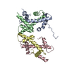
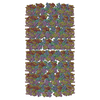
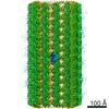
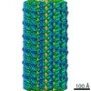
| Title | Iterative Helical Real Space Reconstruction of Histone Octamer Helical Tubes | |||||||||
 Map data Map data | surface rendering of histone octamer tubular crystal | |||||||||
 Sample Sample |
| |||||||||
 Keywords Keywords | histone / octamer / H2A / H2B / H3 / H4 / nucleosome / IHRSR / helical / tubular crystal | |||||||||
| Function / homology |  Function and homology information Function and homology informationPKMTs methylate histone lysines / HDMs demethylate histones / RMTs methylate histone arginines / SUMOylation of chromatin organization proteins / Condensation of Prophase Chromosomes / Nonhomologous End-Joining (NHEJ) / G2/M DNA damage checkpoint / Processing of DNA double-strand break ends / Interleukin-7 signaling / Chromatin modifying enzymes ...PKMTs methylate histone lysines / HDMs demethylate histones / RMTs methylate histone arginines / SUMOylation of chromatin organization proteins / Condensation of Prophase Chromosomes / Nonhomologous End-Joining (NHEJ) / G2/M DNA damage checkpoint / Processing of DNA double-strand break ends / Interleukin-7 signaling / Chromatin modifying enzymes / Factors involved in megakaryocyte development and platelet production / Formation of the beta-catenin:TCF transactivating complex / : / Transcriptional regulation by small RNAs / : / Metalloprotease DUBs / PRC2 methylates histones and DNA / Oxidative Stress Induced Senescence / HATs acetylate histones / Recruitment and ATM-mediated phosphorylation of repair and signaling proteins at DNA double strand breaks / Assembly of the ORC complex at the origin of replication / RNA Polymerase I Promoter Opening / RNA Polymerase I Promoter Escape / RUNX1 regulates genes involved in megakaryocyte differentiation and platelet function / Estrogen-dependent gene expression / Negative Regulation of CDH1 Gene Transcription / MLL4 and MLL3 complexes regulate expression of PPARG target genes in adipogenesis and hepatic steatosis / Regulation of endogenous retroelements by KRAB-ZFP proteins / Regulation of endogenous retroelements by the Human Silencing Hub (HUSH) complex / UCH proteinases / B-WICH complex positively regulates rRNA expression / Ub-specific processing proteases / Deposition of new CENPA-containing nucleosomes at the centromere / structural constituent of chromatin / nucleosome / nucleosome assembly / heterochromatin formation / protein heterodimerization activity / protein-containing complex binding / DNA binding / nucleoplasm / nucleus Similarity search - Function | |||||||||
| Biological species |  | |||||||||
| Method | helical reconstruction / negative staining / Resolution: 20.0 Å | |||||||||
 Authors Authors | Frouws TD / Patterton HG / Sewell BT | |||||||||
 Citation Citation | Journal: Biophys J / Year: 2009 Title: Histone octamer helical tubes suggest that an internucleosomal four-helix bundle stabilizes the chromatin fiber. Authors: Timothy D Frouws / Hugh-G Patterton / Bryan T Sewell /  Abstract: A major question in chromatin involves the exact organization of nucleosomes within the 30-nm chromatin fiber and its structural determinants of assembly. Here we investigate the structure of histone ...A major question in chromatin involves the exact organization of nucleosomes within the 30-nm chromatin fiber and its structural determinants of assembly. Here we investigate the structure of histone octamer helical tubes via the method of iterative helical real-space reconstruction. Accurate placement of the x-ray structure of the histone octamer within the reconstructed density yields a pseudoatomic model for the entire helix, and allows precise identification of molecular interactions between neighboring octamers. One such interaction that would not be obscured by DNA in the nucleosome consists of a twofold symmetric four-helix bundle formed between pairs of H2B-alpha3 and H2B-alphaC helices of neighboring octamers. We believe that this interface can act as an internucleosomal four-helix bundle within the context of the chromatin fiber. The potential relevance of this interface in the folding of the 30-nm chromatin fiber is discussed. | |||||||||
| History |
|
- Structure visualization
| Movie |
Movie viewer |
|---|---|
| Structure viewer | EM map: SurfViewMolmilJmol/JSmol |
| Supplemental images |
 UCSF Chimera
- Downloads & links
-EMDB archive
| Map data | emd_1469.map.gz | 2.4 MB | EMDB map data format | |
|---|---|---|---|---|
| Header (meta data) | emd-1469-v30.xmlemd-1469.xml | 15.9 KB 15.9 KB | Display Display | EMDB header |
| Images |  1469.gif 1469.gif | 109.3 KB | ||
| Archive directory |  http://ftp.pdbj.org/pub/emdb/structures/EMD-1469ftp://ftp.pdbj.org/pub/emdb/structures/EMD-1469 http://ftp.pdbj.org/pub/emdb/structures/EMD-1469ftp://ftp.pdbj.org/pub/emdb/structures/EMD-1469 | HTTPS FTP |
-Related structure data
| Related structure data | 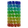 3c9kMC M: atomic model generated by this map C: citing same article ( |
|---|---|
| Similar structure data |


-Links
| EMDB pages | EMDB (EBI/PDBe) / EMDataResource |
|---|---|
| Related items in Molecule of the Month |


-Map
| File | Download / File: emd_1469.map.gz / Format: CCP4 / Size: 7.8 MB / Type: IMAGE STORED AS FLOATING POINT NUMBER (4 BYTES) | ||||||||||||||||||||||||||||||||||||||||||||||||||||||||||||||||||||
|---|---|---|---|---|---|---|---|---|---|---|---|---|---|---|---|---|---|---|---|---|---|---|---|---|---|---|---|---|---|---|---|---|---|---|---|---|---|---|---|---|---|---|---|---|---|---|---|---|---|---|---|---|---|---|---|---|---|---|---|---|---|---|---|---|---|---|---|---|---|
| Annotation | surface rendering of histone octamer tubular crystal | ||||||||||||||||||||||||||||||||||||||||||||||||||||||||||||||||||||
| Projections & slices | Image control
Images are generated by Spider. | ||||||||||||||||||||||||||||||||||||||||||||||||||||||||||||||||||||
| Voxel size | X=Y=Z: 4 Å | ||||||||||||||||||||||||||||||||||||||||||||||||||||||||||||||||||||
| Density |
| ||||||||||||||||||||||||||||||||||||||||||||||||||||||||||||||||||||
| Symmetry | Space group: 1 | ||||||||||||||||||||||||||||||||||||||||||||||||||||||||||||||||||||
| Details | EMDB XML:
CCP4 map header:
| ||||||||||||||||||||||||||||||||||||||||||||||||||||||||||||||||||||
 Z (Sec.)
 Y (Row.)
 X (Col.)


-Supplemental data
- Sample components
-Entire : Histone Octamer Tubular Crystals
| Entire | Name: Histone Octamer Tubular Crystals |
|---|---|
| Components |
|
-Supramolecule #1000: Histone Octamer Tubular Crystals
| Supramolecule | Name: Histone Octamer Tubular Crystals / type: sample / ID: 1000 / Details: crystalline tubes / Oligomeric state: octameric, C11, and helical / Number unique components: 4 |
|---|---|
| Molecular weight | Experimental: 109 KDa / Theoretical: 109 KDa / Method: sequence |
-Macromolecule #1: histone H2A
| Macromolecule | Name: histone H2A / type: protein_or_peptide / ID: 1 / Name.synonym: H2A / Details: Histone octamers purified from chicken blood / Number of copies: 2 / Oligomeric state: octamer / Recombinant expression: No / Database: NCBI |
|---|---|
| Source (natural) | Organism: |
| Molecular weight | Experimental: 14 KDa / Theoretical: 14 KDa |
| Sequence | InterPro: Histone H2A |
-Macromolecule #2: histone H2B
| Macromolecule | Name: histone H2B / type: protein_or_peptide / ID: 2 / Name.synonym: H2B / Details: Histone octamers purified from chicken blood / Number of copies: 2 / Oligomeric state: octamer / Recombinant expression: No / Database: NCBI |
|---|---|
| Source (natural) | Organism: |
| Molecular weight | Experimental: 14 KDa / Theoretical: 14 KDa |
| Sequence | InterPro: Histone H2B |
-Macromolecule #3: histone H3
| Macromolecule | Name: histone H3 / type: protein_or_peptide / ID: 3 / Name.synonym: H3 / Details: Histone octamers purified from chicken blood / Number of copies: 2 / Oligomeric state: octamer / Recombinant expression: No / Database: NCBI |
|---|---|
| Source (natural) | Organism: |
| Molecular weight | Experimental: 15 KDa / Theoretical: 15 KDa |
| Sequence | InterPro: Histone H3/CENP-A |
-Macromolecule #4: histone H4
| Macromolecule | Name: histone H4 / type: protein_or_peptide / ID: 4 / Name.synonym: H4 / Details: Histone octamers purified from chicken blood / Number of copies: 2 / Oligomeric state: octamer / Recombinant expression: No / Database: NCBI |
|---|---|
| Source (natural) | Organism: |
| Molecular weight | Experimental: 11 KDa / Theoretical: 11 KDa |
| Sequence | InterPro: Histone H4 |
-Experimental details
-Structure determination
| Method | negative staining |
|---|---|
 Processing Processing | helical reconstruction |
| Aggregation state | helical array |
-Sample preparation
| Concentration | 2 mg/mL |
|---|---|
| Buffer | pH: 7.4 / Details: 100mM Tris-HCl, 2M NaCl, 40% NH2SO4 |
| Staining | Type: NEGATIVE Details: 5 ul 0.4% Uranyl Acetate is added to 5 ul sample, grid is floated on mixture for 1 min, then washed twice with 10 ul 0.2% Uranyl Acetate drops. |
| Grid | Details: 300 Mesh copper grid |
| Vitrification | Cryogen name: NONE / Instrument: OTHER |
| Details | double dialysis |
- Electron microscopy
| Microscope | ZEISS LEO912 |
|---|---|
| Temperature | Average: 295 K |
| Alignment procedure | Legacy - Astigmatism: corrected at 80,000 times magnification |
| Specialist optics | Energy filter - Name: Omega in-column / Energy filter - Lower energy threshold: 0.0 eV / Energy filter - Upper energy threshold: 15.0 eV |
| Details | Zeiss LEO 912 microscope using low dose technique |
| Image recording | Category: FILM / Film or detector model: KODAK SO-163 FILM / Digitization - Scanner: OTHER / Digitization - Sampling interval: 2 µm / Number real images: 50 / Average electron dose: 100 e/Å2 Details: scanned on Ilford Leafscan at 2A per pixel and binned to 4A per pixel Od range: 1 / Bits/pixel: 16 |
| Tilt angle min | 0 |
| Tilt angle max | 0 |
| Electron beam | Acceleration voltage: 120 kV / Electron source: TUNGSTEN HAIRPIN |
| Electron optics | Illumination mode: FLOOD BEAM / Imaging mode: BRIGHT FIELD / Nominal defocus max: 1.0 µm / Nominal defocus min: 0.8 µm / Nominal magnification: 50000 |
| Sample stage | Specimen holder: Eucentric / Specimen holder model: OTHER |
-Image processing
| Details | Helices segmented into overlapping boxes using Boxer program from EMAN |
|---|---|
| Final reconstruction | Applied symmetry - Helical parameters - Δz: 65.12 Å Applied symmetry - Helical parameters - Δ&Phi: 7.56 ° Applied symmetry - Helical parameters - Axial symmetry: D11 (2x11 fold dihedral) Algorithm: OTHER / Resolution.type: BY AUTHOR / Resolution: 20.0 Å / Resolution method: FSC 0.5 CUT-OFF / Software - Name: SPIDER Details: helical symmetry is searched for and imposed each iteration round on the backprojected volume. |
| Final angle assignment | Details: Single axis tilt geometry. 4.1 degree increment for phi to cover an asymmetric wedge of 32.72 degrees. Therefore 8 reference projections. |
-Atomic model buiding 1
| Initial model | PDB ID: |
|---|---|
| Software | Name: SPIDER |
| Details | Protocol: Rigid body. Because of helical line group, the dyad axis is constrained to lie facing the helical axis and reduces the search to 2D. i.e. to rotations and translations about this dyad. |
| Refinement | Space: REAL / Protocol: RIGID BODY FIT / Target criteria: CC and VdW |
| Output model | PDB-3c9k: |