+ Open data
- Basic information
| Entry | Database: EMDB / ID: EMD-24214 | |||||||||
|---|---|---|---|---|---|---|---|---|---|---|
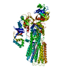
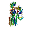
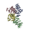
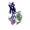
| Title | Cryo-EM structure of ATP13A2 in the BeF-bound E2P-like state | |||||||||
 Map data Map data | Summed, unsharpened map | |||||||||
 Sample Sample |
| |||||||||
 Keywords Keywords | P-type ATPase / P5B-ATPase / polyamine transporter / TRANSPORT PROTEIN | |||||||||
| Function / homology |  Function and homology information Function and homology informationpolyamine transmembrane transporter activity / polyamine transmembrane transport / spermine transmembrane transport / : / ABC-type polyamine transporter activity / regulation of autophagosome size / extracellular exosome biogenesis / regulation of chaperone-mediated autophagy / negative regulation of lysosomal protein catabolic process / P-type ion transporter activity ...polyamine transmembrane transporter activity / polyamine transmembrane transport / spermine transmembrane transport / : / ABC-type polyamine transporter activity / regulation of autophagosome size / extracellular exosome biogenesis / regulation of chaperone-mediated autophagy / negative regulation of lysosomal protein catabolic process / P-type ion transporter activity / regulation of autophagy of mitochondrion / regulation of lysosomal protein catabolic process / intracellular monoatomic cation homeostasis / autophagosome-lysosome fusion / protein localization to lysosome / autophagosome organization / phosphatidic acid binding / positive regulation of exosomal secretion / ATPase-coupled monoatomic cation transmembrane transporter activity / multivesicular body membrane / regulation of protein localization to nucleus / intracellular zinc ion homeostasis / cupric ion binding / phosphatidylinositol-3,5-bisphosphate binding / regulation of mitochondrion organization / Translocases; Catalysing the translocation of other compounds; Linked to the hydrolysis of a nucleoside triphosphate / cellular response to zinc ion / lysosomal transport / regulation of intracellular protein transport / lipid homeostasis / Ion transport by P-type ATPases / autophagosome membrane / cellular response to manganese ion / regulation of macroautophagy / transport vesicle / regulation of neuron apoptotic process / multivesicular body / autophagosome / lysosomal lumen / positive regulation of protein secretion / autophagy / intracellular calcium ion homeostasis / late endosome membrane / late endosome / manganese ion binding / cellular response to oxidative stress / monoatomic ion transmembrane transport / vesicle / intracellular iron ion homeostasis / lysosome / neuron projection / lysosomal membrane / neuronal cell body / positive regulation of gene expression / ATP hydrolysis activity / zinc ion binding / ATP binding / membrane Similarity search - Function | |||||||||
| Biological species |  Homo sapiens (human) Homo sapiens (human) | |||||||||
| Method | single particle reconstruction / cryo EM / Resolution: 2.8 Å | |||||||||
 Authors Authors | Sim SI / Park E | |||||||||
| Funding support |  United States, 1 items United States, 1 items
| |||||||||
 Citation Citation |  Journal: Mol Cell / Year: 2021 Journal: Mol Cell / Year: 2021Title: Structural basis of polyamine transport by human ATP13A2 (PARK9). Authors: Sue Im Sim / Sören von Bülow / Gerhard Hummer / Eunyong Park /   Abstract: Polyamines are small, organic polycations that are ubiquitous and essential to all forms of life. Currently, how polyamines are transported across membranes is not understood. Recent studies have ...Polyamines are small, organic polycations that are ubiquitous and essential to all forms of life. Currently, how polyamines are transported across membranes is not understood. Recent studies have suggested that ATP13A2 and its close homologs, collectively known as P5B-ATPases, are polyamine transporters at endo-/lysosomes. Loss-of-function mutations of ATP13A2 in humans cause hereditary early-onset Parkinson's disease. To understand the polyamine transport mechanism of ATP13A2, we determined high-resolution cryoelectron microscopy (cryo-EM) structures of human ATP13A2 in five distinct conformational intermediates, which together, represent a near-complete transport cycle of ATP13A2. The structural basis of the polyamine specificity was revealed by an endogenous polyamine molecule bound to a narrow, elongated cavity within the transmembrane domain. The structures show an atypical transport path for a water-soluble substrate, in which polyamines may exit within the cytosolic leaflet of the membrane. Our study provides important mechanistic insights into polyamine transport and a framework to understand the functions and mechanisms of P5B-ATPases. | |||||||||
| History |
|
- Structure visualization
| Movie |
 Movie viewer Movie viewer |
|---|---|
| Structure viewer | EM map:  SurfView SurfView Molmil Molmil Jmol/JSmol Jmol/JSmol |
| Supplemental images |
- Downloads & links
-EMDB archive
| Map data |  emd_24214.map.gz emd_24214.map.gz | 62.8 MB |  EMDB map data format EMDB map data format | |
|---|---|---|---|---|
| Header (meta data) |  emd-24214-v30.xml emd-24214-v30.xml emd-24214.xml emd-24214.xml | 25.4 KB 25.4 KB | Display Display |  EMDB header EMDB header |
| FSC (resolution estimation) |  emd_24214_fsc.xml emd_24214_fsc.xml | 11.5 KB | Display |  FSC data file FSC data file |
| Images |  emd_24214.png emd_24214.png | 57.7 KB | ||
| Filedesc metadata |  emd-24214.cif.gz emd-24214.cif.gz | 7.1 KB | ||
| Others |  emd_24214_additional_1.map.gz emd_24214_additional_1.map.gz emd_24214_half_map_1.map.gz emd_24214_half_map_1.map.gz emd_24214_half_map_2.map.gz emd_24214_half_map_2.map.gz | 118.1 MB 115.8 MB 115.8 MB | ||
| Archive directory |  http://ftp.pdbj.org/pub/emdb/structures/EMD-24214 http://ftp.pdbj.org/pub/emdb/structures/EMD-24214 ftp://ftp.pdbj.org/pub/emdb/structures/EMD-24214 ftp://ftp.pdbj.org/pub/emdb/structures/EMD-24214 | HTTPS FTP |
-Validation report
| Summary document |  emd_24214_validation.pdf.gz emd_24214_validation.pdf.gz | 1002.7 KB | Display |  EMDB validaton report EMDB validaton report |
|---|---|---|---|---|
| Full document |  emd_24214_full_validation.pdf.gz emd_24214_full_validation.pdf.gz | 1002.2 KB | Display | |
| Data in XML |  emd_24214_validation.xml.gz emd_24214_validation.xml.gz | 19.1 KB | Display | |
| Data in CIF |  emd_24214_validation.cif.gz emd_24214_validation.cif.gz | 24.7 KB | Display | |
| Arichive directory |  https://ftp.pdbj.org/pub/emdb/validation_reports/EMD-24214 https://ftp.pdbj.org/pub/emdb/validation_reports/EMD-24214 ftp://ftp.pdbj.org/pub/emdb/validation_reports/EMD-24214 ftp://ftp.pdbj.org/pub/emdb/validation_reports/EMD-24214 | HTTPS FTP |
-Related structure data
| Related structure data |  7n70MC  7n72C  7n73C  7n74C  7n75C  7n76C  7n77C  7n78C C: citing same article ( M: atomic model generated by this map |
|---|---|
| Similar structure data |
- Links
Links
| EMDB pages |  EMDB (EBI/PDBe) / EMDB (EBI/PDBe) /  EMDataResource EMDataResource |
|---|---|
| Related items in Molecule of the Month |
- Map
Map
| File |  Download / File: emd_24214.map.gz / Format: CCP4 / Size: 125 MB / Type: IMAGE STORED AS FLOATING POINT NUMBER (4 BYTES) Download / File: emd_24214.map.gz / Format: CCP4 / Size: 125 MB / Type: IMAGE STORED AS FLOATING POINT NUMBER (4 BYTES) | ||||||||||||||||||||||||||||||||||||||||||||||||||||||||||||
|---|---|---|---|---|---|---|---|---|---|---|---|---|---|---|---|---|---|---|---|---|---|---|---|---|---|---|---|---|---|---|---|---|---|---|---|---|---|---|---|---|---|---|---|---|---|---|---|---|---|---|---|---|---|---|---|---|---|---|---|---|---|
| Annotation | Summed, unsharpened map | ||||||||||||||||||||||||||||||||||||||||||||||||||||||||||||
| Projections & slices | Image control
Images are generated by Spider. | ||||||||||||||||||||||||||||||||||||||||||||||||||||||||||||
| Voxel size | X=Y=Z: 0.911 Å | ||||||||||||||||||||||||||||||||||||||||||||||||||||||||||||
| Density |
| ||||||||||||||||||||||||||||||||||||||||||||||||||||||||||||
| Symmetry | Space group: 1 | ||||||||||||||||||||||||||||||||||||||||||||||||||||||||||||
| Details | EMDB XML:
CCP4 map header:
| ||||||||||||||||||||||||||||||||||||||||||||||||||||||||||||
-Supplemental data
-Additional map: Summed, sharpened map
| File | emd_24214_additional_1.map | ||||||||||||
|---|---|---|---|---|---|---|---|---|---|---|---|---|---|
| Annotation | Summed, sharpened map | ||||||||||||
| Projections & Slices |
| ||||||||||||
| Density Histograms |
-Half map: Half map 1
| File | emd_24214_half_map_1.map | ||||||||||||
|---|---|---|---|---|---|---|---|---|---|---|---|---|---|
| Annotation | Half map 1 | ||||||||||||
| Projections & Slices |
| ||||||||||||
| Density Histograms |
-Half map: Half map 2
| File | emd_24214_half_map_2.map | ||||||||||||
|---|---|---|---|---|---|---|---|---|---|---|---|---|---|
| Annotation | Half map 2 | ||||||||||||
| Projections & Slices |
| ||||||||||||
| Density Histograms |
- Sample components
-Entire : Human ATP13A2 complexed with BeF3
| Entire | Name: Human ATP13A2 complexed with BeF3 |
|---|---|
| Components |
|
-Supramolecule #1: Human ATP13A2 complexed with BeF3
| Supramolecule | Name: Human ATP13A2 complexed with BeF3 / type: complex / ID: 1 / Parent: 0 / Macromolecule list: #1 |
|---|---|
| Source (natural) | Organism:  Homo sapiens (human) Homo sapiens (human) |
-Macromolecule #1: Isoform 3 of Polyamine-transporting ATPase 13A2
| Macromolecule | Name: Isoform 3 of Polyamine-transporting ATPase 13A2 / type: protein_or_peptide / ID: 1 / Number of copies: 1 / Enantiomer: LEVO EC number: Translocases; Catalysing the translocation of other compounds; Linked to the hydrolysis of a nucleoside triphosphate |
|---|---|
| Source (natural) | Organism:  Homo sapiens (human) Homo sapiens (human) |
| Molecular weight | Theoretical: 128.444469 KDa |
| Recombinant expression | Organism:  |
| Sequence | String: MSADSSPLVG STPTGYGTLT IGTSIDPLSS SVSSVRLSGY CGSPWRVIGY HVVVWMMAGI PLLLFRWKPL WGVRLRLRPC NLAHAETLV IEIRDKEDSS WQLFTVQVQT EAIGEGSLEP SPQSQAEDGR SQAAVGAVPE GAWKDTAQLH KSEEAKRVLR Y YLFQGQRY ...String: MSADSSPLVG STPTGYGTLT IGTSIDPLSS SVSSVRLSGY CGSPWRVIGY HVVVWMMAGI PLLLFRWKPL WGVRLRLRPC NLAHAETLV IEIRDKEDSS WQLFTVQVQT EAIGEGSLEP SPQSQAEDGR SQAAVGAVPE GAWKDTAQLH KSEEAKRVLR Y YLFQGQRY IWIETQQAFY QVSLLDHGRS CDDVHRSRHG LSLQDQMVRK AIYGPNVISI PVKSYPQLLV DEALNPYYGF QA FSIALWL ADHYYWYALC IFLISSISIC LSLYKTRKQS QTLRDMVKLS MRVCVCRPGG EEEWVDSSEL VPGDCLVLPQ EGG LMPCDA ALVAGECMVN ESSLTGESIP VLKTALPEGL GPYCAETHRR HTLFCGTLIL QARAYVGPHV LAVVTRTGFC TAKG GLVSS ILHPRPINFK FYKHSMKFVA ALSVLALLGT IYSIFILYRN RVPLNEIVIR ALDLVTVVVP PALPAAMTVC TLYAQ SRLR RQGIFCIHPL RINLGGKLQL VCFDKTGTLT EDGLDVMGVV PLKGQAFLPL VPEPRRLPVG PLLRALATCH ALSRLQ DTP VGDPMDLKMV ESTGWVLEEE PAADSAFGTQ VLAVMRPPLW EPQLQAMEEP PVPVSVLHRF PFSSALQRMS VVVAWPG AT QPEAYVKGSP ELVAGLCNPE TVPTDFAQML QSYTAAGYRV VALASKPLPT VPSLEAAQQL TRDTVEGDLS LLGLLVMR N LLKPQTTPVI QALRRTRIRA VMVTGDNLQT AVTVARGCGM VAPQEHLIIV HATHPERGQP ASLEFLPMES PTAVNGVKD PDQAASYTVE PDPRSRHLAL SGPTFGIIVK HFPKLLPKVL VQGTVFARMA PEQKTELVCE LQKLQYCVGM CGDGANDCGA LKAADVGIS LSQAEASVVS PFTSSMASIE CVPMVIREGR CSLDTSFSVF KYMALYSLTQ FISVLILYTI NTNLGDLQFL A IDLVITTT VAVLMSRTGP ALVLGRVRPP GALLSVPVLS SLLLQMVLVT GVQLGGYFLT LAQPWFVPLN RTVAAPDNLP NY ENTVVFS LSSFQYLILA AAVSKGAPFR RPLYTNVPFL VALALLSSVL VGLVLVPGLL QGPLALRNIT DTGFKLLLLG LVT LNFVGA FMLESVLDQC LPACLRRLRP KRASKKRFKQ LERELAEQPW PPLPAGPLR UniProtKB: Polyamine-transporting ATPase 13A2 |
-Macromolecule #2: BERYLLIUM TRIFLUORIDE ION
| Macromolecule | Name: BERYLLIUM TRIFLUORIDE ION / type: ligand / ID: 2 / Number of copies: 1 / Formula: BEF |
|---|---|
| Molecular weight | Theoretical: 66.007 Da |
| Chemical component information | 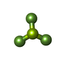 ChemComp-BEF: |
-Macromolecule #3: MAGNESIUM ION
| Macromolecule | Name: MAGNESIUM ION / type: ligand / ID: 3 / Number of copies: 1 / Formula: MG |
|---|---|
| Molecular weight | Theoretical: 24.305 Da |
-Macromolecule #4: CHOLESTEROL HEMISUCCINATE
| Macromolecule | Name: CHOLESTEROL HEMISUCCINATE / type: ligand / ID: 4 / Number of copies: 1 / Formula: Y01 |
|---|---|
| Molecular weight | Theoretical: 486.726 Da |
| Chemical component information |  ChemComp-Y01: |
-Macromolecule #5: DODECYL-BETA-D-MALTOSIDE
| Macromolecule | Name: DODECYL-BETA-D-MALTOSIDE / type: ligand / ID: 5 / Number of copies: 1 / Formula: LMT |
|---|---|
| Molecular weight | Theoretical: 510.615 Da |
| Chemical component information |  ChemComp-LMT: |
-Experimental details
-Structure determination
| Method | cryo EM |
|---|---|
 Processing Processing | single particle reconstruction |
| Aggregation state | particle |
- Sample preparation
| Buffer | pH: 7.5 |
|---|---|
| Grid | Model: Quantifoil R1.2/1.3 / Material: GOLD / Mesh: 400 / Support film - Material: CARBON / Support film - topology: HOLEY / Pretreatment - Type: GLOW DISCHARGE / Pretreatment - Time: 40 sec. / Pretreatment - Atmosphere: AIR / Pretreatment - Pressure: 0.039 kPa |
| Vitrification | Cryogen name: ETHANE / Chamber humidity: 100 % / Chamber temperature: 277 K / Instrument: FEI VITROBOT MARK IV |
- Electron microscopy
| Microscope | FEI TITAN KRIOS |
|---|---|
| Image recording | Film or detector model: GATAN K3 (6k x 4k) / Average electron dose: 50.0 e/Å2 |
| Electron beam | Acceleration voltage: 300 kV / Electron source:  FIELD EMISSION GUN FIELD EMISSION GUN |
| Electron optics | Illumination mode: FLOOD BEAM / Imaging mode: BRIGHT FIELD / Cs: 2.7 mm |
| Experimental equipment |  Model: Titan Krios / Image courtesy: FEI Company |
 Movie
Movie Controller







 Z (Sec.)
 Y (Row.)
 X (Col.)


