+ Open data
- Basic information
| Entry | Database: EMDB / ID: EMD-22413 | |||||||||
|---|---|---|---|---|---|---|---|---|---|---|
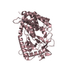
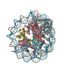
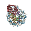
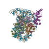
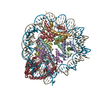
| Title | 1:1 cGAS(mask)-NCP map | |||||||||
 Map data Map data | 1:! cGAS(mask)-nucleosome map | |||||||||
 Sample Sample |
| |||||||||
| Method | single particle reconstruction / cryo EM / Resolution: 3.9 Å | |||||||||
 Authors Authors | Boyer JA / Spangler CJ / Strauss JD / McGinty RK / Zhang Q | |||||||||
| Funding support |  United States, 2 items United States, 2 items
| |||||||||
 Citation Citation |  Journal: Science / Year: 2020 Journal: Science / Year: 2020Title: Structural basis of nucleosome-dependent cGAS inhibition. Authors: Joshua A Boyer / Cathy J Spangler / Joshua D Strauss / Andrew P Cesmat / Pengda Liu / Robert K McGinty / Qi Zhang /  Abstract: Cyclic guanosine monophosphate (GMP)-adenosine monophosphate (AMP) synthase (cGAS) recognizes cytosolic foreign or damaged DNA to activate the innate immune response to infection, inflammatory ...Cyclic guanosine monophosphate (GMP)-adenosine monophosphate (AMP) synthase (cGAS) recognizes cytosolic foreign or damaged DNA to activate the innate immune response to infection, inflammatory diseases, and cancer. By contrast, cGAS reactivity against self-DNA in the nucleus is suppressed by chromatin tethering. We report a 3.3-angstrom-resolution cryo-electron microscopy structure of cGAS in complex with the nucleosome core particle. The structure reveals that cGAS uses two conserved arginines to anchor to the nucleosome acidic patch. The nucleosome-binding interface exclusively occupies the strong double-stranded DNA (dsDNA)-binding surface on cGAS and sterically prevents cGAS from oligomerizing into the functionally active 2:2 cGAS-dsDNA state. These findings provide a structural basis for how cGAS maintains an inhibited state in the nucleus and further exemplify the role of the nucleosome in regulating diverse nuclear protein functions. | |||||||||
| History |
|
- Structure visualization
| Movie |
 Movie viewer Movie viewer |
|---|---|
| Structure viewer | EM map:  SurfView SurfView Molmil Molmil Jmol/JSmol Jmol/JSmol |
| Supplemental images |
- Downloads & links
-EMDB archive
| Map data |  emd_22413.map.gz emd_22413.map.gz | 9 MB |  EMDB map data format EMDB map data format | |
|---|---|---|---|---|
| Header (meta data) |  emd-22413-v30.xml emd-22413-v30.xml emd-22413.xml emd-22413.xml | 15.2 KB 15.2 KB | Display Display |  EMDB header EMDB header |
| FSC (resolution estimation) |  emd_22413_fsc.xml emd_22413_fsc.xml | 11.6 KB | Display |  FSC data file FSC data file |
| Images |  emd_22413.png emd_22413.png | 79.1 KB | ||
| Others |  emd_22413_additional.map.gz emd_22413_additional.map.gz emd_22413_additional_1.map.gz emd_22413_additional_1.map.gz | 73.9 MB 73.9 MB | ||
| Archive directory |  http://ftp.pdbj.org/pub/emdb/structures/EMD-22413 http://ftp.pdbj.org/pub/emdb/structures/EMD-22413 ftp://ftp.pdbj.org/pub/emdb/structures/EMD-22413 ftp://ftp.pdbj.org/pub/emdb/structures/EMD-22413 | HTTPS FTP |
-Validation report
| Summary document |  emd_22413_validation.pdf.gz emd_22413_validation.pdf.gz | 79 KB | Display |  EMDB validaton report EMDB validaton report |
|---|---|---|---|---|
| Full document |  emd_22413_full_validation.pdf.gz emd_22413_full_validation.pdf.gz | 78.1 KB | Display | |
| Data in XML |  emd_22413_validation.xml.gz emd_22413_validation.xml.gz | 493 B | Display | |
| Arichive directory |  https://ftp.pdbj.org/pub/emdb/validation_reports/EMD-22413 https://ftp.pdbj.org/pub/emdb/validation_reports/EMD-22413 ftp://ftp.pdbj.org/pub/emdb/validation_reports/EMD-22413 ftp://ftp.pdbj.org/pub/emdb/validation_reports/EMD-22413 | HTTPS FTP |
-Related structure data
- Links
Links
| EMDB pages |  EMDB (EBI/PDBe) / EMDB (EBI/PDBe) /  EMDataResource EMDataResource |
|---|
- Map
Map
| File |  Download / File: emd_22413.map.gz / Format: CCP4 / Size: 137.1 MB / Type: IMAGE STORED AS FLOATING POINT NUMBER (4 BYTES) Download / File: emd_22413.map.gz / Format: CCP4 / Size: 137.1 MB / Type: IMAGE STORED AS FLOATING POINT NUMBER (4 BYTES) | ||||||||||||||||||||||||||||||||||||||||||||||||||||||||||||||||||||
|---|---|---|---|---|---|---|---|---|---|---|---|---|---|---|---|---|---|---|---|---|---|---|---|---|---|---|---|---|---|---|---|---|---|---|---|---|---|---|---|---|---|---|---|---|---|---|---|---|---|---|---|---|---|---|---|---|---|---|---|---|---|---|---|---|---|---|---|---|---|
| Annotation | 1:! cGAS(mask)-nucleosome map | ||||||||||||||||||||||||||||||||||||||||||||||||||||||||||||||||||||
| Projections & slices | Image control
Images are generated by Spider. | ||||||||||||||||||||||||||||||||||||||||||||||||||||||||||||||||||||
| Voxel size | X=Y=Z: 0.91 Å | ||||||||||||||||||||||||||||||||||||||||||||||||||||||||||||||||||||
| Density |
| ||||||||||||||||||||||||||||||||||||||||||||||||||||||||||||||||||||
| Symmetry | Space group: 1 | ||||||||||||||||||||||||||||||||||||||||||||||||||||||||||||||||||||
| Details | EMDB XML:
CCP4 map header:
| ||||||||||||||||||||||||||||||||||||||||||||||||||||||||||||||||||||
-Supplemental data
-Additional map: 1:1 cGAS-nucleosome composite map
| File | emd_22413_additional.map | ||||||||||||
|---|---|---|---|---|---|---|---|---|---|---|---|---|---|
| Annotation | 1:1 cGAS-nucleosome composite map | ||||||||||||
| Projections & Slices |
| ||||||||||||
| Density Histograms |
-Additional map: 1:1 cGAS-nucleosome composite map
| File | emd_22413_additional_1.map | ||||||||||||
|---|---|---|---|---|---|---|---|---|---|---|---|---|---|
| Annotation | 1:1 cGAS-nucleosome composite map | ||||||||||||
| Projections & Slices |
| ||||||||||||
| Density Histograms |
- Sample components
-Entire : 1:1 cGAS-nucleosome complex
| Entire | Name: 1:1 cGAS-nucleosome complex |
|---|---|
| Components |
|
-Supramolecule #1: 1:1 cGAS-nucleosome complex
| Supramolecule | Name: 1:1 cGAS-nucleosome complex / type: complex / ID: 1 / Parent: 0 / Macromolecule list: #1-#7 / Details: mouse cGAS bound to the nucleosome in a 1:1 ratio |
|---|---|
| Molecular weight | Theoretical: 250 KDa |
-Experimental details
-Structure determination
| Method | cryo EM |
|---|---|
 Processing Processing | single particle reconstruction |
| Aggregation state | particle |
- Sample preparation
| Concentration | 1 mg/mL | ||||||||||||
|---|---|---|---|---|---|---|---|---|---|---|---|---|---|
| Buffer | pH: 7.5 Component:
| ||||||||||||
| Grid | Model: Quantifoil / Material: COPPER / Mesh: 400 / Support film - Material: CARBON / Support film - topology: HOLEY / Pretreatment - Type: GLOW DISCHARGE / Details: Instrument: Pelco easiGlow | ||||||||||||
| Vitrification | Cryogen name: ETHANE / Chamber humidity: 95 % / Chamber temperature: 293 K / Instrument: FEI VITROBOT MARK IV |
- Electron microscopy
| Microscope | FEI TALOS ARCTICA |
|---|---|
| Image recording | Film or detector model: GATAN K3 (6k x 4k) / Number grids imaged: 1 / Number real images: 2100 / Average electron dose: 53.0 e/Å2 |
| Electron beam | Acceleration voltage: 200 kV / Electron source:  FIELD EMISSION GUN FIELD EMISSION GUN |
| Electron optics | Illumination mode: FLOOD BEAM / Imaging mode: BRIGHT FIELD |
| Experimental equipment |  Model: Talos Arctica / Image courtesy: FEI Company |
 Movie
Movie Controller




 Z (Sec.)
 Y (Row.)
 X (Col.)


