 Movie
Movie Controller


+ Open data

- Basic information
| Entry | Database: EMDB / ID: EMD-20309 | |||||||||
|---|---|---|---|---|---|---|---|---|---|---|
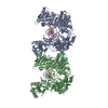
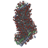
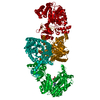
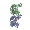
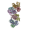
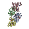
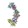
| Title | cryoEM structure of yeast glucokinase filament | |||||||||
 Map data Map data | cryoEM structure of yeast glucokinase filament | |||||||||
 Sample Sample |
| |||||||||
 Keywords Keywords | filament / TRANSFERASE | |||||||||
| Function / homology |  Function and homology information Function and homology informationSynthesis of GDP-mannose / glucokinase / Glycolysis / fructokinase activity / glucokinase activity / mannose metabolic process / Regulation of Glucokinase by Glucokinase Regulatory Protein / glucose 6-phosphate metabolic process / D-glucose binding / : ...Synthesis of GDP-mannose / glucokinase / Glycolysis / fructokinase activity / glucokinase activity / mannose metabolic process / Regulation of Glucokinase by Glucokinase Regulatory Protein / glucose 6-phosphate metabolic process / D-glucose binding / : / Neutrophil degranulation / intracellular glucose homeostasis / glycolytic process / glucose metabolic process / mitochondrion / ATP binding / plasma membrane / cytosol Similarity search - Function | |||||||||
| Biological species |  | |||||||||
| Method | helical reconstruction / cryo EM / Resolution: 3.8 Å | |||||||||
 Authors Authors | Lynch EM / Dosey AM | |||||||||
 Citation Citation | Journal: Science / Year: 2020 Title: Polymerization in the actin ATPase clan regulates hexokinase activity in yeast. Authors: Patrick R Stoddard / Eric M Lynch / Daniel P Farrell / Annie M Dosey / Frank DiMaio / Tom A Williams / Justin M Kollman / Andrew W Murray / Ethan C Garner /   Abstract: The actin fold is found in cytoskeletal polymers, chaperones, and various metabolic enzymes. Many actin-fold proteins, such as the carbohydrate kinases, do not polymerize. We found that Glk1, a ...The actin fold is found in cytoskeletal polymers, chaperones, and various metabolic enzymes. Many actin-fold proteins, such as the carbohydrate kinases, do not polymerize. We found that Glk1, a glucokinase, forms two-stranded filaments with ultrastructure that is distinct from that of cytoskeletal polymers. In cells, Glk1 polymerized upon sugar addition and depolymerized upon sugar withdrawal. Polymerization inhibits enzymatic activity; the Glk1 monomer-polymer equilibrium sets a maximum rate of glucose phosphorylation regardless of Glk1 concentration. A mutation that eliminated Glk1 polymerization alleviated concentration-dependent enzyme inhibition. Yeast containing nonpolymerizing Glk1 were less fit when growing on sugars and more likely to die when refed glucose. Glk1 polymerization arose independently from other actin-related filaments and may allow yeast to rapidly modulate glucokinase activity as nutrient availability changes. | |||||||||
| History |
|
- Structure visualization
| Movie |
Movie viewer |
|---|---|
| Structure viewer | EM map: SurfViewMolmilJmol/JSmol |
| Supplemental images |

- Downloads & links
-EMDB archive
| Map data | emd_20309.map.gz | 7.4 MB | EMDB map data format | |
|---|---|---|---|---|
| Header (meta data) | emd-20309-v30.xmlemd-20309.xml | 11.1 KB 11.1 KB | Display Display | EMDB header |
| Images |  emd_20309.png emd_20309.png | 137.1 KB | ||
| Filedesc metadata | emd-20309.cif.gz | 5.5 KB | ||
| Archive directory |  http://ftp.pdbj.org/pub/emdb/structures/EMD-20309ftp://ftp.pdbj.org/pub/emdb/structures/EMD-20309 http://ftp.pdbj.org/pub/emdb/structures/EMD-20309ftp://ftp.pdbj.org/pub/emdb/structures/EMD-20309 | HTTPS FTP |
-Related structure data
| Related structure data | 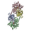 6pdtMC 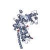 6p4xC M: atomic model generated by this map C: citing same article ( |
|---|---|
| Similar structure data |

-Links
| EMDB pages | EMDB (EBI/PDBe) / EMDataResource |
|---|---|
| Related items in Molecule of the Month |


-Map
| File | Download / File: emd_20309.map.gz / Format: CCP4 / Size: 125 MB / Type: IMAGE STORED AS FLOATING POINT NUMBER (4 BYTES) | ||||||||||||||||||||||||||||||||||||||||||||||||||||||||||||||||||||
|---|---|---|---|---|---|---|---|---|---|---|---|---|---|---|---|---|---|---|---|---|---|---|---|---|---|---|---|---|---|---|---|---|---|---|---|---|---|---|---|---|---|---|---|---|---|---|---|---|---|---|---|---|---|---|---|---|---|---|---|---|---|---|---|---|---|---|---|---|---|
| Annotation | cryoEM structure of yeast glucokinase filament | ||||||||||||||||||||||||||||||||||||||||||||||||||||||||||||||||||||
| Projections & slices | Image control
Images are generated by Spider. | ||||||||||||||||||||||||||||||||||||||||||||||||||||||||||||||||||||
| Voxel size | X=Y=Z: 1.05 Å | ||||||||||||||||||||||||||||||||||||||||||||||||||||||||||||||||||||
| Density |
| ||||||||||||||||||||||||||||||||||||||||||||||||||||||||||||||||||||
| Symmetry | Space group: 1 | ||||||||||||||||||||||||||||||||||||||||||||||||||||||||||||||||||||
| Details | EMDB XML:
CCP4 map header:
| ||||||||||||||||||||||||||||||||||||||||||||||||||||||||||||||||||||
 Z (Sec.)
 Y (Row.)
 X (Col.)


-Supplemental data
- Sample components
-Entire : glucokinase-1
| Entire | Name: glucokinase-1 |
|---|---|
| Components |
|
-Supramolecule #1: glucokinase-1
| Supramolecule | Name: glucokinase-1 / type: complex / ID: 1 / Parent: 0 / Macromolecule list: #1 |
|---|---|
| Source (natural) | Organism: |
-Macromolecule #1: Glucokinase-1
| Macromolecule | Name: Glucokinase-1 / type: protein_or_peptide / ID: 1 / Number of copies: 4 / Enantiomer: LEVO / EC number: glucokinase |
|---|---|
| Source (natural) | Organism: Strain: ATCC 204508 / S288c |
| Molecular weight | Theoretical: 55.446258 KDa |
| Recombinant expression | Organism:  |
| Sequence | String: MSFDDLHKAT ERAVIQAVDQ ICDDFEVTPE KLDELTAYFI EQMEKGLAPP KEGHTLASDK GLPMIPAFVT GSPNGTERGV LLAADLGGT NFRICSVNLH GDHTFSMEQM KSKIPDDLLD DENVTSDDLF GFLARRTLAF MKKYHPDELA KGKDAKPMKL G FTFSYPVD ...String: MSFDDLHKAT ERAVIQAVDQ ICDDFEVTPE KLDELTAYFI EQMEKGLAPP KEGHTLASDK GLPMIPAFVT GSPNGTERGV LLAADLGGT NFRICSVNLH GDHTFSMEQM KSKIPDDLLD DENVTSDDLF GFLARRTLAF MKKYHPDELA KGKDAKPMKL G FTFSYPVD QTSLNSGTLI RWTKGFRIAD TVGKDVVQLY QEQLSAQGMP MIKVVALTND TVGTYLSHCY TSDNTDSMTS GE ISEPVIG CIFGTGTNGC YMEEINKITK LPQELRDKLI KEGKTHMIIN VEWGSFDNEL KHLPTTKYDV VIDQKLSTNP GFH LFEKRV SGMFLGEVLR NILVDLHSQG LLLQQYRSKE QLPRHLTTPF QLSSEVLSHI EIDDSTGLRE TELSLLQSLR LPTT PTERV QIQKLVRAIS RRSAYLAAVP LAAILIKTNA LNKRYHGEVE IGCDGSVVEY YPGFRSMLRH ALALSPLGAE GERKV HLKI AKDGSGVGAA LCALVA UniProtKB: Glucokinase-1 |
-Macromolecule #2: alpha-D-glucopyranose
| Macromolecule | Name: alpha-D-glucopyranose / type: ligand / ID: 2 / Number of copies: 4 / Formula: GLC |
|---|---|
| Molecular weight | Theoretical: 180.156 Da |
| Chemical component information |  ChemComp-GLC: |
-Macromolecule #3: MAGNESIUM ION
| Macromolecule | Name: MAGNESIUM ION / type: ligand / ID: 3 / Number of copies: 4 / Formula: MG |
|---|---|
| Molecular weight | Theoretical: 24.305 Da |
-Macromolecule #4: ADENOSINE-5'-TRIPHOSPHATE
| Macromolecule | Name: ADENOSINE-5'-TRIPHOSPHATE / type: ligand / ID: 4 / Number of copies: 4 / Formula: ATP |
|---|---|
| Molecular weight | Theoretical: 507.181 Da |
| Chemical component information |  ChemComp-ATP: |
-Experimental details
-Structure determination
| Method | cryo EM |
|---|---|
 Processing Processing | helical reconstruction |
| Aggregation state | filament |
-Sample preparation
| Buffer | pH: 7.5 |
|---|---|
| Vitrification | Cryogen name: ETHANE / Chamber humidity: 100 % |
- Electron microscopy
| Microscope | FEI TITAN KRIOS |
|---|---|
| Image recording | Film or detector model: GATAN K2 SUMMIT (4k x 4k) / Detector mode: SUPER-RESOLUTION / Average electron dose: 90.0 e/Å2 |
| Electron beam | Acceleration voltage: 300 kV / Electron source:  FIELD EMISSION GUN FIELD EMISSION GUN |
| Electron optics | Illumination mode: FLOOD BEAM / Imaging mode: BRIGHT FIELD |
| Experimental equipment |  Model: Titan Krios / Image courtesy: FEI Company |
-Image processing
| Final reconstruction | Applied symmetry - Helical parameters - Δz: 60.1 Å Applied symmetry - Helical parameters - Δ&Phi: 120.4 ° Applied symmetry - Helical parameters - Axial symmetry: D1 (2x1 fold dihedral) Resolution.type: BY AUTHOR / Resolution: 3.8 Å / Resolution method: FSC 0.143 CUT-OFF / Software - Name: RELION / Number images used: 56778 |
|---|---|
| Startup model | Type of model: OTHER |
| Final angle assignment | Type: NOT APPLICABLE / Software - Name: RELION |
