 Movie
Movie Controller


+ Open data

- Basic information
| Entry | Database: EMDB / ID: EMD-20091 | |||||||||
|---|---|---|---|---|---|---|---|---|---|---|
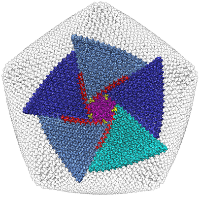
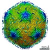
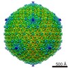
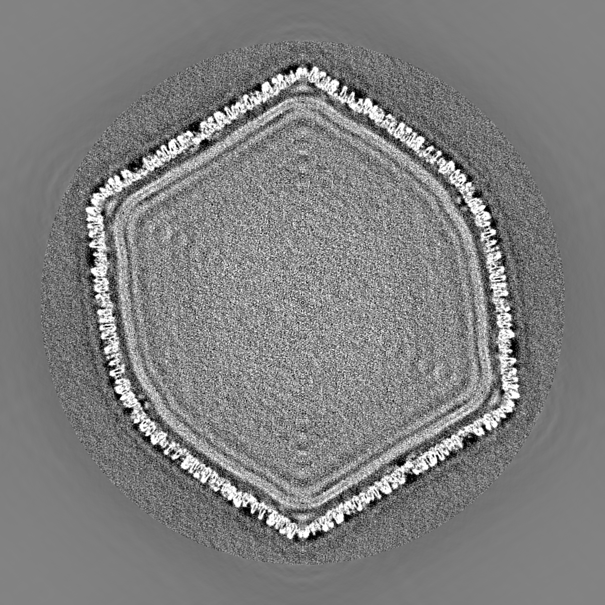
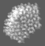
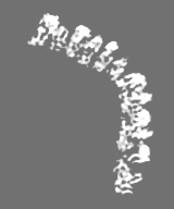
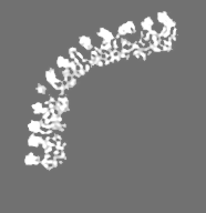
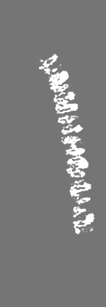
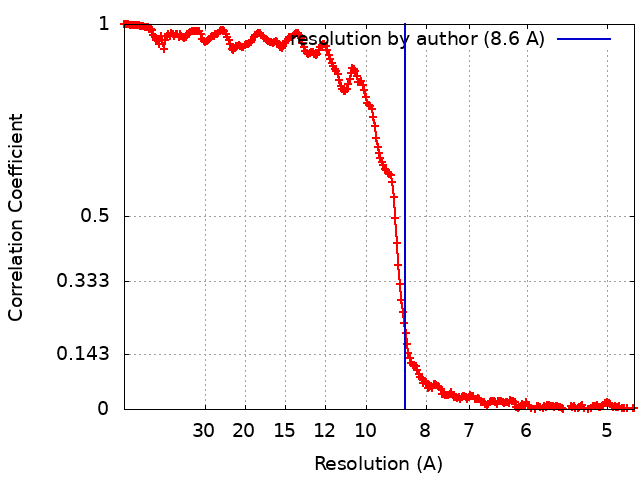
| Title | Singapore Grouper Iridovirus (SGIV) at 8.6 Angstrom Resolution | |||||||||
 Map data Map data | Independent reconstruction half of the micrographs | |||||||||
 Sample Sample |
| |||||||||
 Keywords Keywords | icosahedral / trimers / outer coat / anchor proteins / zip proteins / VIRAL PROTEIN | |||||||||
| Function / homology | Major capsid protein, N-terminal / Major capsid protein N-terminus / Major capsid protein, C-terminal / Major capsid protein, C-terminal domain superfamily / Large eukaryotic DNA virus major capsid protein / Group II dsDNA virus coat/capsid protein / viral capsid / structural molecule activity / Major capsid protein Function and homology information Function and homology information | |||||||||
| Biological species |  Singapore grouper iridovirus Singapore grouper iridovirus | |||||||||
| Method | single particle reconstruction / cryo EM / Resolution: 8.6 Å | |||||||||
 Authors Authors | Pintilie G / Chen D-H | |||||||||
| Funding support |  United States, 2 items United States, 2 items
| |||||||||
 Citation Citation | Journal: Structure / Year: 2019 Title: Segmentation and Comparative Modeling in an 8.6-Å Cryo-EM Map of the Singapore Grouper Iridovirus. Authors: Grigore Pintilie / Dong-Hua Chen / Bich Ngoc Tran / Joanita Jakana / Jinlu Wu / Choy Leong Hew / Wah Chiu /  Abstract: SGIV, or Singapore grouper iridovirus, is a large double-stranded DNA virus, reaching a diameter of 220 nm and packaging a genome of 140 kb. We present a 3D cryoelectron microscopy (cryo-EM) ...SGIV, or Singapore grouper iridovirus, is a large double-stranded DNA virus, reaching a diameter of 220 nm and packaging a genome of 140 kb. We present a 3D cryoelectron microscopy (cryo-EM) icosahedral reconstruction of SGIV determined at 8.6-Å resolution. It reveals several layers including a T = 247 icosahedral outer coat, anchor proteins, a lipid bilayer, and the encapsidated DNA. A new segmentation tool, iSeg, was applied to extract these layers from the reconstructed map. The outer coat was further segmented into major and minor capsid proteins. None of the proteins extracted by segmentation have known atomic structures. We generated models for the major coat protein using three comparative modeling tools, and evaluated each model using the cryo-EM map. Our analysis reveals a new architecture in the Iridoviridae family of viruses. It shares similarities with others in the same family, e.g., Chilo iridescent virus, but also shows new features of the major and minor capsid proteins. | |||||||||
| History |
|
- Structure visualization
| Movie |
Movie viewer |
|---|---|
| Structure viewer | EM map: SurfViewMolmilJmol/JSmol |
| Supplemental images |
- Downloads & links
-EMDB archive
 emd_20091.png
-Related structure data
| Related structure data | 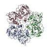 6ojnMC M: atomic model generated by this map C: citing same article ( |
|---|---|
| Similar structure data |


- Links
Links
| EMDB pages | EMDB (EBI/PDBe) / EMDataResource |
|---|---|
| Related items in Molecule of the Month |


-Map
| File | Download / File: emd_20091.map.gz / Format: CCP4 / Size: 6.4 GB / Type: IMAGE STORED AS FLOATING POINT NUMBER (4 BYTES) | ||||||||||||||||||||||||||||||||||||||||||||||||||||||||||||
|---|---|---|---|---|---|---|---|---|---|---|---|---|---|---|---|---|---|---|---|---|---|---|---|---|---|---|---|---|---|---|---|---|---|---|---|---|---|---|---|---|---|---|---|---|---|---|---|---|---|---|---|---|---|---|---|---|---|---|---|---|---|
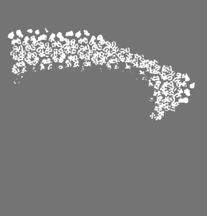
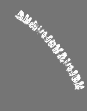
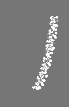
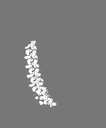
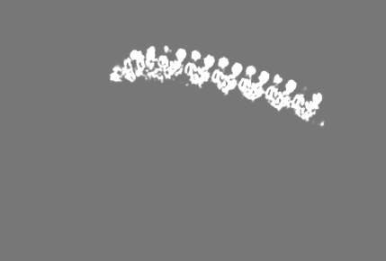
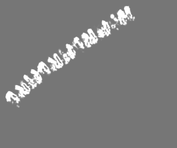
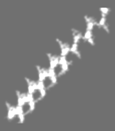
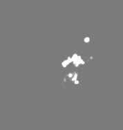
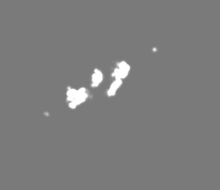
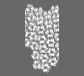
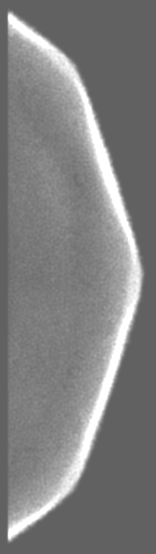
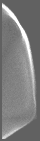
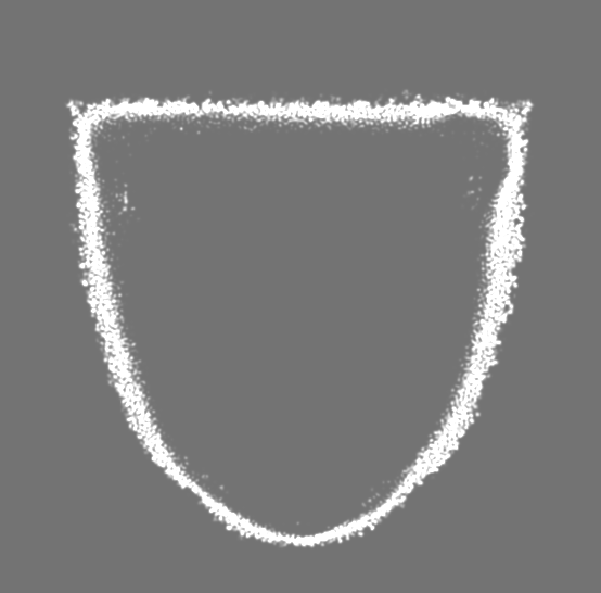
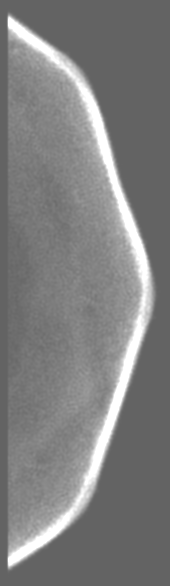
| Annotation | Independent reconstruction half of the micrographs | ||||||||||||||||||||||||||||||||||||||||||||||||||||||||||||
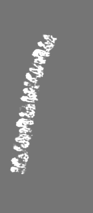
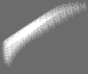
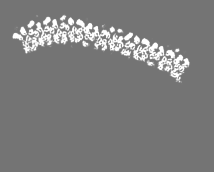
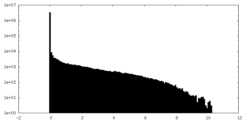
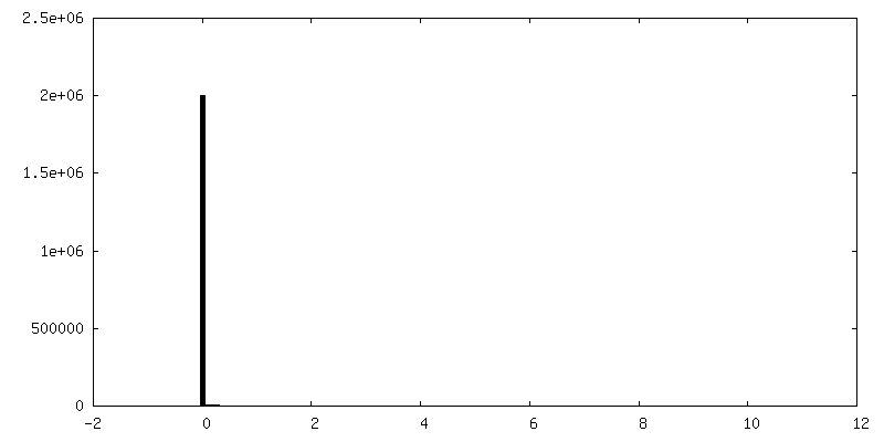
| Projections & slices | Image control
Images are generated by Spider. | ||||||||||||||||||||||||||||||||||||||||||||||||||||||||||||
| Voxel size | X=Y=Z: 2.37 Å | ||||||||||||||||||||||||||||||||||||||||||||||||||||||||||||
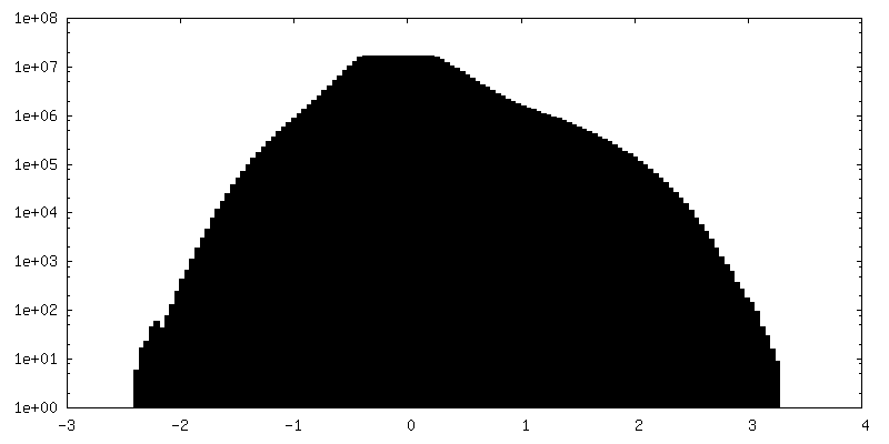
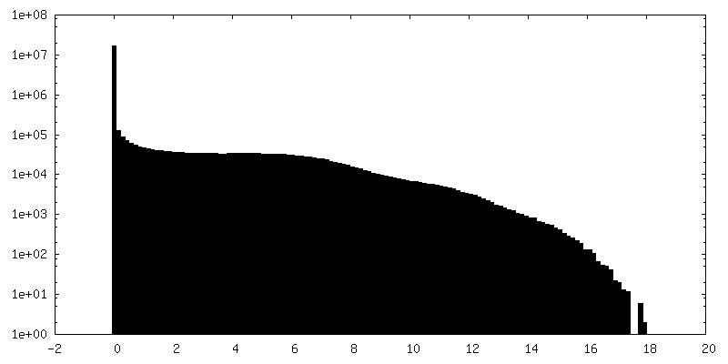
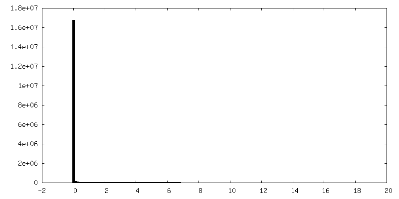
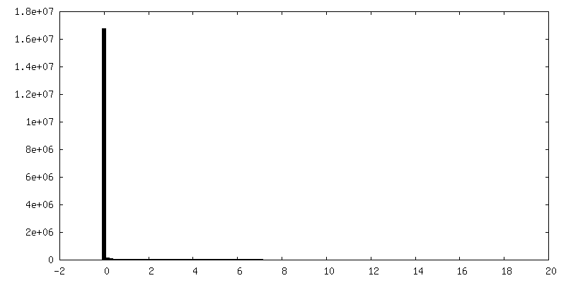
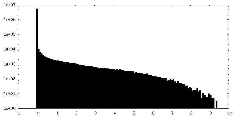
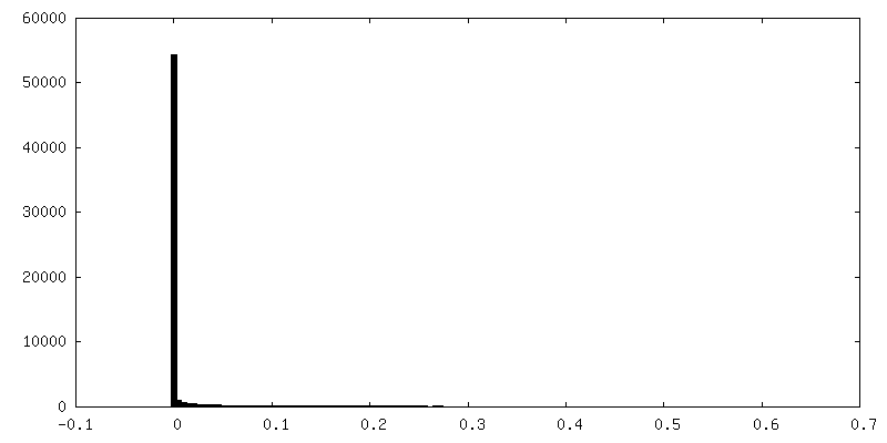
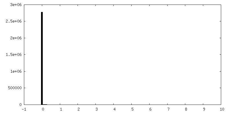
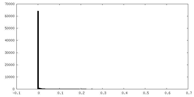
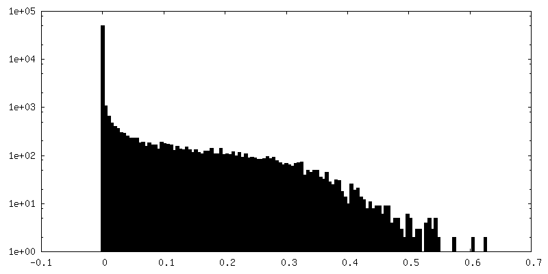
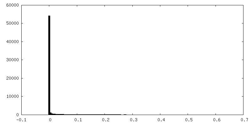
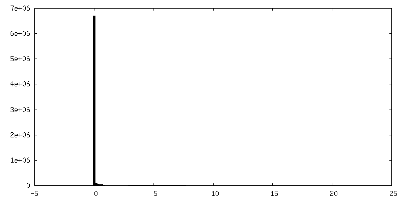
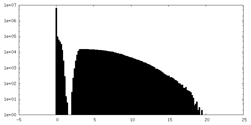
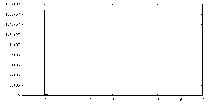
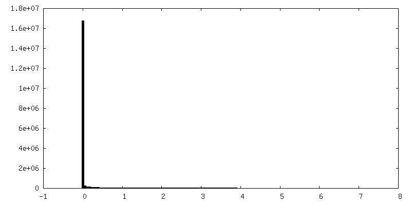
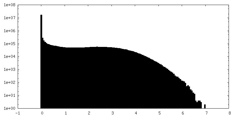
| Density |
| ||||||||||||||||||||||||||||||||||||||||||||||||||||||||||||
| Symmetry | Space group: 1 | ||||||||||||||||||||||||||||||||||||||||||||||||||||||||||||
| Details | EMDB XML:
CCP4 map header:
| ||||||||||||||||||||||||||||||||||||||||||||||||||||||||||||
 Z (Sec.)
 Y (Row.)
 X (Col.)


-Supplemental data
+Additional map: Independent reconstruction other half of the micrographs


+Additional map: A pentasymmetron


+Additional map: Averaged densities from trimers from a trisymmetron within...


+Additional map: Trisymmetron 1 of 5 around a pentasymmetron


+Additional map: Trisymmetron 2 of 5 around a pentasymmetron


+Additional map: Trisymmetron 3 of 5 around a pentasymmetron


+Supplemental map: emd 20091 additional 15.map


+Additional map: Trisymmetron 5 of 5 around a pentasymmetron


+Additional map: Zip proteins 1


+Additional map: Zip proteins 2


+Additional map: Zip proteins 3


+Additional map: Anchor protein 1


+Additional map: Zip proteins 4


+Additional map: Zip proteins 5


+Additional map: Anchor protein 2


+Additional map: Anchor protein 3


+Additional map: Anchor protein 4


+Additional map: Anchor protein 5


+Additional map: An asymmetric unit from the icosahedrally-symmetric map


+Additional map: Inner half of the bilayer lipid membrane


+Additional map: Outer half of the bilayer lipid membrane


- Sample components
-Entire : Singapore grouper iridovirus
| Entire | Name: Singapore grouper iridovirus |
|---|---|
| Components |
|
-Supramolecule #1: Singapore grouper iridovirus
| Supramolecule | Name: Singapore grouper iridovirus / type: virus / ID: 1 / Parent: 0 / Macromolecule list: all / NCBI-ID: 262968 / Sci species name: Singapore grouper iridovirus / Virus type: VIRION / Virus isolate: SPECIES / Virus enveloped: Yes / Virus empty: No |
|---|---|
| Host (natural) | Organism:  Epinephelus tauvina (greasy grouper) Epinephelus tauvina (greasy grouper) |
| Virus shell | Shell ID: 1 / Name: Outer coat / Diameter: 2200.0 Å / T number (triangulation number): 247 |
-Macromolecule #1: Major capsid protein
| Macromolecule | Name: Major capsid protein / type: protein_or_peptide / ID: 1 / Number of copies: 3 / Enantiomer: LEVO |
|---|---|
| Source (natural) | Organism: Singapore grouper iridovirus |
| Molecular weight | Theoretical: 50.573141 KDa |
| Sequence | String: MTCTTGAGVT SGFIDLATYD NLDRALYGGK DATTYFIKEH YPVGWFTKLP TMATRVSGNP AFGQEFSVGV PRSGDYVLNA WLTLKTPEI KLLETNRLGA NGTVRWTKNL MHNAVEHASL TFNDICAQQF NTAYLDAWTQ FNMCEGKRIG YDNMIGNTSD M TNPTPAQG ...String: MTCTTGAGVT SGFIDLATYD NLDRALYGGK DATTYFIKEH YPVGWFTKLP TMATRVSGNP AFGQEFSVGV PRSGDYVLNA WLTLKTPEI KLLETNRLGA NGTVRWTKNL MHNAVEHASL TFNDICAQQF NTAYLDAWTQ FNMCEGKRIG YDNMIGNTSD M TNPTPAQG QDGARTLPSK NLVLPLPFFF SRDCGLALPT VVLPYNEIRI NIKLRSLQEL LVFQNKDTGN VIPISATDIA GG LADTVEA YVYMTVGLVS NVERCAMAGT VRDMVVEQMQ AAPTHIVNPQ NTNNVHVDMR FSHAVKALFF MVQNVTYKSV GSN YTCVTP VNGPGNTVME PAMSVDPIKS ASLTYENTTR LANMGVEYYS LVQPWYFSAS IPVYTGYHMY SYALNVGSVH PSGS TNYGR LTNASITVTM SPESVVAAAG GGNNNSGYNE PQRFALVVIA VNHNVIRIMN GSMGFPIL UniProtKB: Major capsid protein |
-Experimental details
-Structure determination
| Method | cryo EM |
|---|---|
 Processing Processing | single particle reconstruction |
| Aggregation state | particle |
-Sample preparation
| Buffer | pH: 7.4 |
|---|---|
| Grid | Model: Quantifoil R2/1 |
| Vitrification | Cryogen name: ETHANE / Chamber humidity: 100 % / Instrument: FEI VITROBOT MARK IV / Details: Blotted for 1 second before plunging.. |
- Electron microscopy
| Microscope | FEI TITAN KRIOS |
|---|---|
| Image recording | Film or detector model: GATAN ULTRASCAN 4000 (4k x 4k) / Average exposure time: 1.0 sec. / Average electron dose: 25.0 e/Å2 |
| Electron beam | Acceleration voltage: 300 kV / Electron source:  FIELD EMISSION GUN FIELD EMISSION GUN |
| Electron optics | C2 aperture diameter: 70.0 µm / Calibrated magnification: 63290 / Illumination mode: FLOOD BEAM / Imaging mode: BRIGHT FIELD / Cs: 2.7 mm / Nominal defocus max: 3.0 µm / Nominal defocus min: 1.5 µm |
| Sample stage | Specimen holder model: FEI TITAN KRIOS AUTOGRID HOLDER / Cooling holder cryogen: NITROGEN |
| Experimental equipment |  Model: Titan Krios / Image courtesy: FEI Company |
+Image processing
-Atomic model buiding 1
| Initial model | PDB ID:  1m3y Chain - Source name: PDB / Chain - Initial model type: experimental model |
|---|---|
| Refinement | Space: REAL / Protocol: RIGID BODY FIT / Target criteria: Z-score of Cross Correlation scores |
| Output model | PDB-6ojn: |
