- PDB-6ojn: Comparative Model of SGIV Major Coat Protein (MCP) Trimer Based o... -
+
Open data
ID or keywords:
Loading...
-
Basic information
Entry
Database: PDB / ID: 6ojn
Title
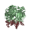
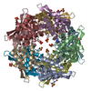
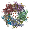
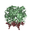
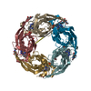
Comparative Model of SGIV Major Coat Protein (MCP) Trimer Based on Cryo-EM Map
Components
Major capsid protein
Keywords
VIRAL PROTEIN / icosahedral / trimers / outer coat / anchor proteins / zip proteins
Function / homology
Major capsid protein, N-terminal / Major capsid protein N-terminus / Major capsid protein, C-terminal / Major capsid protein, C-terminal domain superfamily / Large eukaryotic DNA virus major capsid protein / Group II dsDNA virus coat/capsid protein / viral capsid / structural molecule activity / Major capsid protein
Function and homology information
Biological species
Singapore grouper iridovirus
Method
ELECTRON MICROSCOPY / single particle reconstruction / cryo EM / Resolution: 8.6 Å
National Institutes of Health/National Human Genome Research Institute (NIH/NHGRI)
P41GM103832
United States
National Institutes of Health/National Human Genome Research Institute (NIH/NHGRI)
R01GM079429
United States
Citation
Journal: Structure / Year: 2019 Title: Segmentation and Comparative Modeling in an 8.6-Å Cryo-EM Map of the Singapore Grouper Iridovirus. Authors: Grigore Pintilie / Dong-Hua Chen / Bich Ngoc Tran / Joanita Jakana / Jinlu Wu / Choy Leong Hew / Wah Chiu / Abstract: SGIV, or Singapore grouper iridovirus, is a large double-stranded DNA virus, reaching a diameter of 220 nm and packaging a genome of 140 kb. We present a 3D cryoelectron microscopy (cryo-EM) ...SGIV, or Singapore grouper iridovirus, is a large double-stranded DNA virus, reaching a diameter of 220 nm and packaging a genome of 140 kb. We present a 3D cryoelectron microscopy (cryo-EM) icosahedral reconstruction of SGIV determined at 8.6-Å resolution. It reveals several layers including a T = 247 icosahedral outer coat, anchor proteins, a lipid bilayer, and the encapsidated DNA. A new segmentation tool, iSeg, was applied to extract these layers from the reconstructed map. The outer coat was further segmented into major and minor capsid proteins. None of the proteins extracted by segmentation have known atomic structures. We generated models for the major coat protein using three comparative modeling tools, and evaluated each model using the cryo-EM map. Our analysis reveals a new architecture in the Iridoviridae family of viruses. It shares similarities with others in the same family, e.g., Chilo iridescent virus, but also shows new features of the major and minor capsid proteins.
In the structure databanks used in Yorodumi, some data are registered as the other names, "COVID-19 virus" and "2019-nCoV". Here are the details of the virus and the list of structure data.
Jan 31, 2019. EMDB accession codes are about to change! (news from PDBe EMDB page)
EMDB accession codes are about to change! (news from PDBe EMDB page)
The allocation of 4 digits for EMDB accession codes will soon come to an end. Whilst these codes will remain in use, new EMDB accession codes will include an additional digit and will expand incrementally as the available range of codes is exhausted. The current 4-digit format prefixed with “EMD-” (i.e. EMD-XXXX) will advance to a 5-digit format (i.e. EMD-XXXXX), and so on. It is currently estimated that the 4-digit codes will be depleted around Spring 2019, at which point the 5-digit format will come into force.
The EM Navigator/Yorodumi systems omit the EMD- prefix.
Related info.:Q: What is EMD? / ID/Accession-code notation in Yorodumi/EM Navigator
Yorodumi is a browser for structure data from EMDB, PDB, SASBDB, etc.
This page is also the successor to EM Navigator detail page, and also detail information page/front-end page for Omokage search.
The word "yorodu" (or yorozu) is an old Japanese word meaning "ten thousand". "mi" (miru) is to see.
Related info.:EMDB / PDB / SASBDB / Comparison of 3 databanks / Yorodumi Search / Aug 31, 2016. New EM Navigator & Yorodumi / Yorodumi Papers / Jmol/JSmol / Function and homology information / Changes in new EM Navigator and Yorodumi
 Movie
Movie Controller

 Yorodumi
 Open data
 Basic information
 Components
 Keywords
 Function and homology information
 Singapore grouper iridovirus
 Authors
 United States, 2items
  Citation
 Structure visualization
 Downloads & links
 Other downloads


 PDBj
PDBj

 Assembly
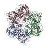
 Sample preparation
 Electron microscopy imaging
 FIELD EMISSION GUN / Accelerating voltage: 300 kV / Illumination mode: FLOOD BEAM
 Processing
