Movie
Movie Controller

[English] 日本語
 Yorodumi
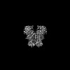
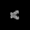
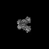
- EMDB-15916: Cryo-EM structure of Ca2+-bound mTMEM16F N562A mutant in Digitoni... -
+ Open data

- Basic information
| Entry |  | |||||||||
|---|---|---|---|---|---|---|---|---|---|---|
| Title | Cryo-EM structure of Ca2+-bound mTMEM16F N562A mutant in Digitonin closed/closed | |||||||||
 Map data Map data | ||||||||||
 Sample Sample |
| |||||||||
 Keywords Keywords | Membrane Protein / Lipid Transport / Lipid Scramblase / Ion Channel / Blood Coagulation / Membrane Fusion | |||||||||
| Function / homology |  Function and homology information Function and homology informationcalcium activated galactosylceramide scrambling / phosphatidylserine exposure on blood platelet / calcium activated phosphatidylserine scrambling / calcium activated phosphatidylcholine scrambling / calcium activated phospholipid scrambling / positive regulation of potassium ion export across plasma membrane / positive regulation of monoatomic ion transmembrane transport / activation of blood coagulation via clotting cascade / purinergic nucleotide receptor signaling pathway / phospholipid scramblase activity ...calcium activated galactosylceramide scrambling / phosphatidylserine exposure on blood platelet / calcium activated phosphatidylserine scrambling / calcium activated phosphatidylcholine scrambling / calcium activated phospholipid scrambling / positive regulation of potassium ion export across plasma membrane / positive regulation of monoatomic ion transmembrane transport / activation of blood coagulation via clotting cascade / purinergic nucleotide receptor signaling pathway / phospholipid scramblase activity / bone mineralization involved in bone maturation / cholinergic synapse / intracellularly calcium-gated chloride channel activity / pore complex assembly / negative regulation of cell volume / plasma membrane phospholipid scrambling / voltage-gated monoatomic ion channel activity / bleb assembly / positive regulation of phagocytosis, engulfment / Stimuli-sensing channels / voltage-gated chloride channel activity / calcium-activated cation channel activity / positive regulation of monocyte chemotaxis / dendritic cell chemotaxis / chloride transport / phospholipid translocation / chloride channel activity / regulation of postsynaptic membrane potential / positive regulation of endothelial cell apoptotic process / positive regulation of bone mineralization / chloride channel complex / Neutrophil degranulation / chloride transmembrane transport / sodium ion transmembrane transport / synaptic membrane / calcium ion transmembrane transport / blood coagulation / positive regulation of apoptotic process / protein homodimerization activity / metal ion binding / identical protein binding / plasma membrane / cytosol Similarity search - Function | |||||||||
| Biological species |  | |||||||||
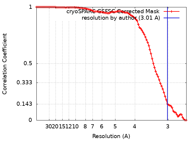
| Method | single particle reconstruction / cryo EM / Resolution: 3.01 Å | |||||||||
 Authors Authors | Arndt M / Alvadia C / Straub MS / Clerico-Mosina V / Paulino C / Dutzler R | |||||||||
| Funding support | European Union, 1 items
| |||||||||
 Citation Citation | Journal: Nat Commun / Year: 2022 Title: Structural basis for the activation of the lipid scramblase TMEM16F. Authors: Melanie Arndt / Carolina Alvadia / Monique S Straub / Vanessa Clerico Mosina / Cristina Paulino / Raimund Dutzler /   Abstract: TMEM16F, a member of the conserved TMEM16 family, plays a central role in the initiation of blood coagulation and the fusion of trophoblasts. The protein mediates passive ion and lipid transport in ...TMEM16F, a member of the conserved TMEM16 family, plays a central role in the initiation of blood coagulation and the fusion of trophoblasts. The protein mediates passive ion and lipid transport in response to an increase in intracellular Ca. However, the mechanism of how the protein facilitates both processes has remained elusive. Here we investigate the basis for TMEM16F activation. In a screen of residues lining the proposed site of conduction, we identify mutants with strongly activating phenotype. Structures of these mutants determined herein by cryo-electron microscopy show major rearrangements leading to the exposure of hydrophilic patches to the membrane, whose distortion facilitates lipid diffusion. The concomitant opening of a pore promotes ion conduction in the same protein conformation. Our work has revealed a mechanism that is distinct for this branch of the family and that will aid the development of a specific pharmacology for a promising drug target. | |||||||||
| History |
|
- Structure visualization
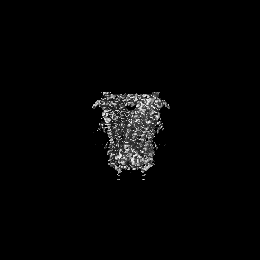
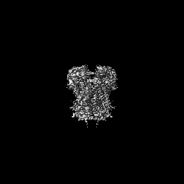
| Supplemental images |
|---|
- Downloads & links
-EMDB archive
| Map data | emd_15916.map.gz | 33.9 MB | EMDB map data format | |
|---|---|---|---|---|
| Header (meta data) | emd-15916-v30.xmlemd-15916.xml | 16.5 KB 16.5 KB | Display Display | EMDB header |
| FSC (resolution estimation) | emd_15916_fsc.xml | 9.6 KB | Display | FSC data file |
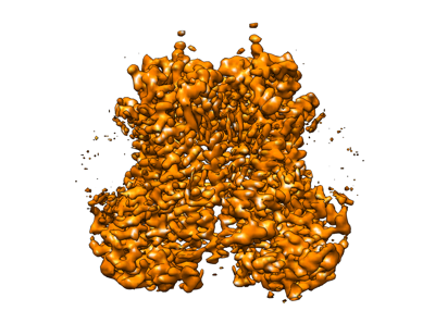
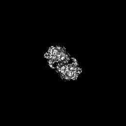
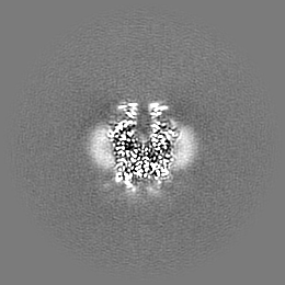
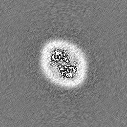
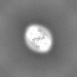
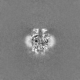
| Images |  emd_15916.png emd_15916.png | 99.9 KB | ||
| Filedesc metadata | emd-15916.cif.gz | 6.4 KB | ||
| Others | emd_15916_half_map_1.map.gzemd_15916_half_map_2.map.gz | 62.1 MB 62.1 MB | ||
| Archive directory |  http://ftp.pdbj.org/pub/emdb/structures/EMD-15916ftp://ftp.pdbj.org/pub/emdb/structures/EMD-15916 http://ftp.pdbj.org/pub/emdb/structures/EMD-15916ftp://ftp.pdbj.org/pub/emdb/structures/EMD-15916 | HTTPS FTP |
-Related structure data
| Related structure data |  8b8kMC 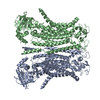 8b8gC  8b8jC 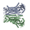 8b8mC  8b8qC 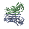 8bc0C  8bc1C M: atomic model generated by this map C: citing same article ( |
|---|---|
| Similar structure data |


-Links
| EMDB pages | EMDB (EBI/PDBe) / EMDataResource |
|---|---|
| Related items in Molecule of the Month |


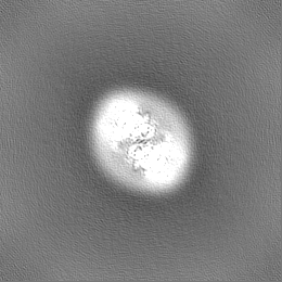
-Map
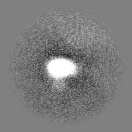
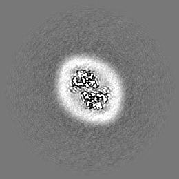
| File | Download / File: emd_15916.map.gz / Format: CCP4 / Size: 67 MB / Type: IMAGE STORED AS FLOATING POINT NUMBER (4 BYTES) | ||||||||||||||||||||||||||||||||||||
|---|---|---|---|---|---|---|---|---|---|---|---|---|---|---|---|---|---|---|---|---|---|---|---|---|---|---|---|---|---|---|---|---|---|---|---|---|---|
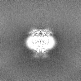
| Projections & slices | Image control
Images are generated by Spider. | ||||||||||||||||||||||||||||||||||||
| Voxel size | X=Y=Z: 1.302 Å | ||||||||||||||||||||||||||||||||||||
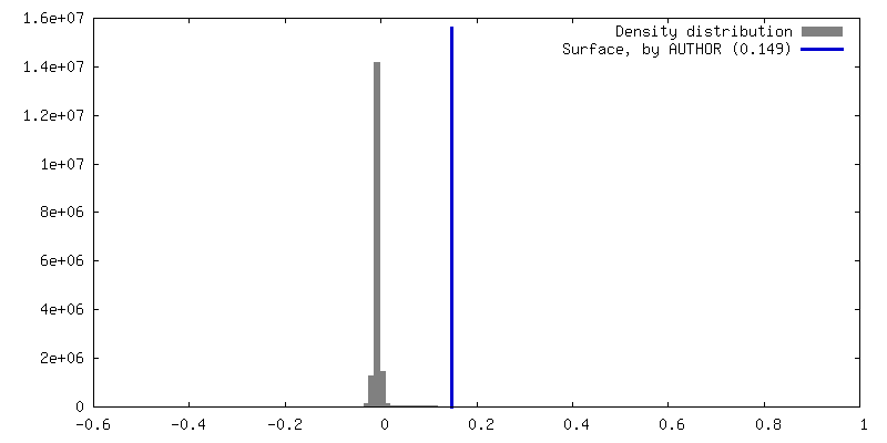
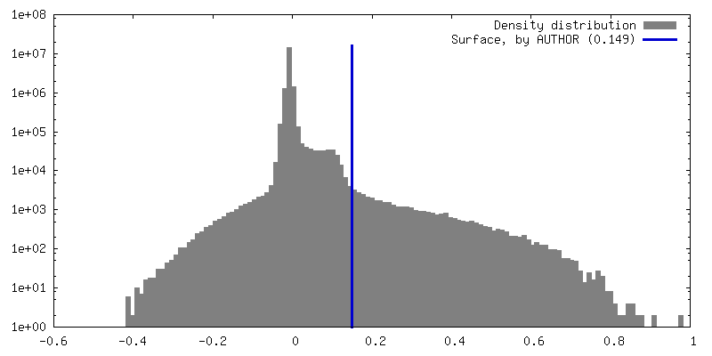
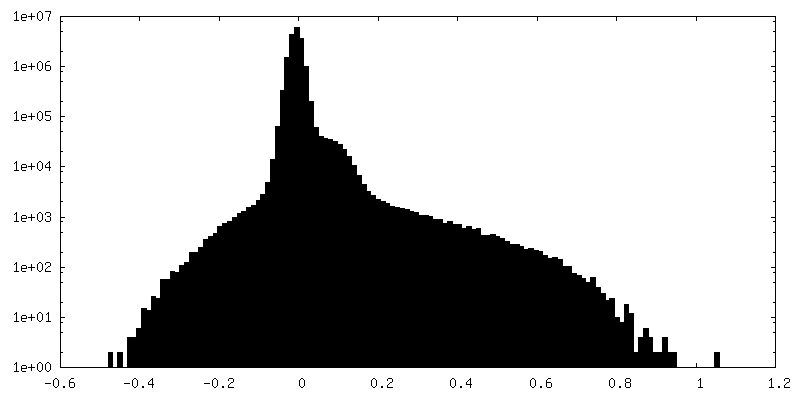
| Density |
| ||||||||||||||||||||||||||||||||||||
| Symmetry | Space group: 1 | ||||||||||||||||||||||||||||||||||||
| Details | EMDB XML:
|
 Z (Sec.)
 Y (Row.)
 X (Col.)


-Supplemental data
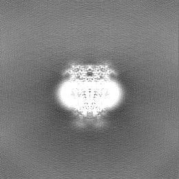
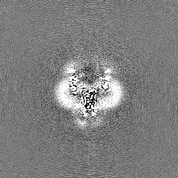
-Half map: #2
| File | emd_15916_half_map_1.map | ||||||||||||
|---|---|---|---|---|---|---|---|---|---|---|---|---|---|
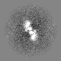
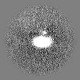
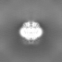
| Projections & Slices |
| ||||||||||||
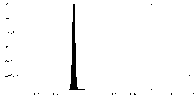
| Density Histograms |
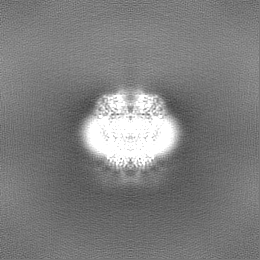
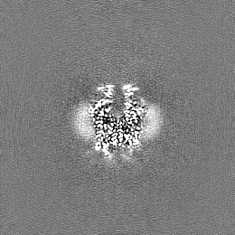

-Half map: #1
| File | emd_15916_half_map_2.map | ||||||||||||
|---|---|---|---|---|---|---|---|---|---|---|---|---|---|
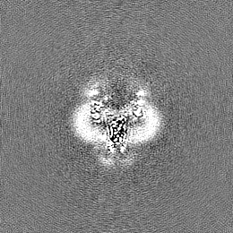
| Projections & Slices |
| ||||||||||||
| Density Histograms |


- Sample components
-Entire : mTMEM16F Ca2+-bound
| Entire | Name: mTMEM16F Ca2+-bound |
|---|---|
| Components |
|
-Supramolecule #1: mTMEM16F Ca2+-bound
| Supramolecule | Name: mTMEM16F Ca2+-bound / type: complex / ID: 1 / Parent: 0 / Macromolecule list: #1 |
|---|---|
| Source (natural) | Organism: |
-Macromolecule #1: Anoctamin-6
| Macromolecule | Name: Anoctamin-6 / type: protein_or_peptide / ID: 1 / Number of copies: 2 / Enantiomer: LEVO |
|---|---|
| Source (natural) | Organism: |
| Molecular weight | Theoretical: 113.420602 KDa |
| Recombinant expression | Organism:  Homo sapiens (human) Homo sapiens (human) |
| Sequence | String: MQMMTRKVLL NMELEEDDDE DGDIVLENFD QTIVCPTFGS LENQQDFRTP EFEEFNGKPD SLFFTDGQRR IDFILVYEDE SKKENNKKG TNEKQKRKRQ AYESNLICHG LQLEATRSVS DDKLVFVKVH APWEVLCTYA EIMHIKLPLK PNDLKTRSPF G NLNWFTKV ...String: MQMMTRKVLL NMELEEDDDE DGDIVLENFD QTIVCPTFGS LENQQDFRTP EFEEFNGKPD SLFFTDGQRR IDFILVYEDE SKKENNKKG TNEKQKRKRQ AYESNLICHG LQLEATRSVS DDKLVFVKVH APWEVLCTYA EIMHIKLPLK PNDLKTRSPF G NLNWFTKV LRVNESVIKP EQEFFTAPFE KSRMNDFYIL DRDSFFNPAT RSRIVYFILS RVKYQVMNNV NKFGINRLVS SG IYKAAFP LHDCRFNYES EDISCPSERY LLYREWAHPR SIYKKQPLDL IRKYYGEKIG IYFAWLGYYT QMLLLAAVVG VAC FLYGYL DQDNCTWSKE VCDPDIGGQI LMCPQCDRLC PFWRLNITCE SSKKLCIFDS FGTLIFAVFM GVWVTLFLEF WKRR QAELE YEWDTVELQQ EEQARPEYEA QCNHVVINEI TQEEERIPFT TCGKCIRVTL CASAVFFWIL LIIASVIGII VYRLS VFIV FSTTLPKNPN GTDPIQKYLT PQMATSITAS IISFIIIMIL NTIYEKVAIM ITNFELPRTQ TDYENSLTMK MFLFQF VAY YSSCFYIAFF KGKFVGYPGD PVYLLGKYRS EECDPGGCLL ELTTQLTIIM GGKAIWNNIQ EVLLPWVMNL IGRYKRV SG SEKITPRWEQ DYHLQPMGKL GLFYEYLEMI IQFGFVTLFV ASFPLAPLLA LVNNILEIRV DAWKLTTQFR RMVPEKAQ D IGAWQPIMQG IAILAVVTNA MIIAFTSDMI PRLVYYWSFS IPPYGDHTYY TMDGYINNTL SVFNITDFKN TDKENPYIG LGNYTLCRYR DFRNPPGHPQ EYKHNIYYWH VIAAKLAFII VMEHIIYSVK FFISYAIPDV SKITKSKIKR EKYLTQKLLH ESHLKDLTK NMGIIAERIG GTVDNSVRPK LEALEVLFQG PQGTEQKLIS EEDLRGASMD EKTTGWRGGH VVEGLAGELE Q LRARLEHH PQGQREP UniProtKB: Anoctamin-6 |
-Macromolecule #2: CALCIUM ION
| Macromolecule | Name: CALCIUM ION / type: ligand / ID: 2 / Number of copies: 6 / Formula: CA |
|---|---|
| Molecular weight | Theoretical: 40.078 Da |
-Macromolecule #3: 1,2-DIDECANOYL-SN-GLYCERO-3-PHOSPHOCHOLINE
| Macromolecule | Name: 1,2-DIDECANOYL-SN-GLYCERO-3-PHOSPHOCHOLINE / type: ligand / ID: 3 / Number of copies: 2 / Formula: P1O |
|---|---|
| Molecular weight | Theoretical: 566.728 Da |
| Chemical component information |  ChemComp-P1O: |
-Experimental details
-Structure determination
| Method | cryo EM |
|---|---|
 Processing Processing | single particle reconstruction |
| Aggregation state | particle |
-Sample preparation
| Buffer | pH: 7.5 / Details: 20mM HEPES 150mM NaCl 2mM EGTA 0.1% Digitonin |
|---|---|
| Grid | Model: Quantifoil R1.2/1.3 / Material: GOLD / Mesh: 200 / Pretreatment - Type: GLOW DISCHARGE / Pretreatment - Time: 30 sec. |
| Vitrification | Cryogen name: ETHANE-PROPANE / Chamber humidity: 100 % / Chamber temperature: 277 K / Instrument: FEI VITROBOT MARK IV / Details: 2mM free Ca2+ added shortly before freezing.. |
- Electron microscopy
| Microscope | FEI TITAN KRIOS |
|---|---|
| Image recording | Film or detector model: GATAN K3 BIOQUANTUM (6k x 4k) / Average electron dose: 62.4 e/Å2 |
| Electron beam | Acceleration voltage: 300 kV / Electron source:  FIELD EMISSION GUN FIELD EMISSION GUN |
| Electron optics | Illumination mode: FLOOD BEAM / Imaging mode: BRIGHT FIELD / Nominal defocus max: 2.4 µm / Nominal defocus min: 1.0 µm |
| Sample stage | Specimen holder model: FEI TITAN KRIOS AUTOGRID HOLDER / Cooling holder cryogen: NITROGEN |
| Experimental equipment |  Model: Titan Krios / Image courtesy: FEI Company |