Loading
PDBj
MenuPDBj@FacebookPDBj@TwitterPDBj@YouTubewwPDB FoundationwwPDB
RCSB PDBPDBeBMRBAdv. SearchSearch help
Search by PDB author
6ET6
DownloadVisualize
BU of 6et6 by Molmil
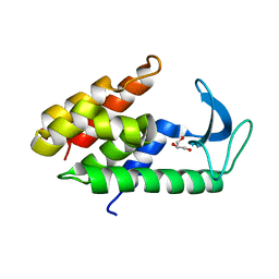
Crystal structure of muramidase from Acinetobacter baumannii AB 5075UW prophage
Descriptor: GLYCEROL, Lysozyme, SULFATE ION
Authors:Boyko, K.M, Nikolaeva, A.Y, Sykilinda, N.N, Shneider, M.M, Miroshnikov, K.A, Popov, V.O.
Deposit date:2017-10-25
Release date:2018-09-05
Last modified:2024-01-17
Method:X-RAY DIFFRACTION (1.2 Å)
Cite:Structure of anAcinetobacterBroad-Range Prophage Endolysin Reveals a C-Terminal alpha-Helix with the Proposed Role in Activity against Live Bacterial Cells.
Viruses, 10, 2018
4GBF
DownloadVisualize
BU of 4gbf by Molmil
Crystal structure of the C-terminal domain of gp131 from bacteriophage phiKZ
Descriptor: CHLORIDE ION, MAGNESIUM ION, PHIKZ131
Authors:Sycheva, L.V, Shneider, M.M, Leiman, P.G.
Deposit date:2012-07-27
Release date:2012-10-17
Last modified:2024-02-28
Method:X-RAY DIFFRACTION (1.949 Å)
Cite:Crystal structure and location of gp131 in the bacteriophage phiKZ virion.
Virology, 434, 2012

218853

PDB entries from 2024-04-24

PDB statisticsPDBj update infoContact PDBjnumon