5HSH
 
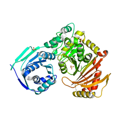 | |
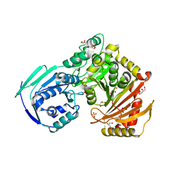
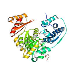
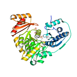
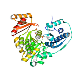
7S77
 | | Crystal structure of the G391V variant of human PGM-1 | | Descriptor: | Phosphoglucomutase-1, SULFATE ION | | Authors: | Stiers, K.M, Beamer, L.J. | | Deposit date: | 2021-09-15 | | Release date: | 2022-05-04 | | Last modified: | 2023-10-18 | | Method: | X-RAY DIFFRACTION (2.8 Å) | | Cite: | Effects of the T337M and G391V disease-related variants on human phosphoglucomutase 1: structural disruptions large and small.
Acta Crystallogr.,Sect.F, 78, 2022
|
|
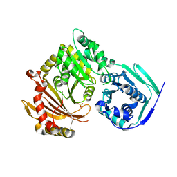
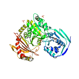
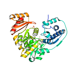
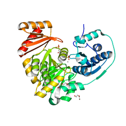
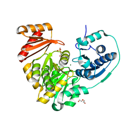
7S0W
 | | Crystal structure of the T337M variant of human PGM-1 | | Descriptor: | COBALT (II) ION, GLYCEROL, Phosphoglucomutase-1, ... | | Authors: | Stiers, K.M, Beamer, L.J. | | Deposit date: | 2021-08-31 | | Release date: | 2022-05-04 | | Last modified: | 2023-10-18 | | Method: | X-RAY DIFFRACTION (2.5 Å) | | Cite: | Effects of the T337M and G391V disease-related variants on human phosphoglucomutase 1: structural disruptions large and small.
Acta Crystallogr.,Sect.F, 78, 2022
|
|
6UIQ
 | |
5VBI
 | |
5VG7
 | |
5VIN
 | |
5VEC
 | |
6NOL
 | |
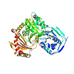
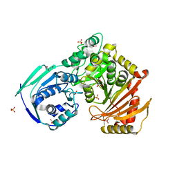
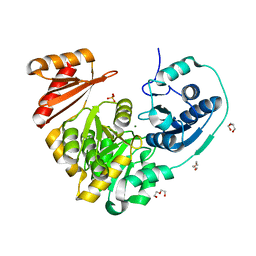
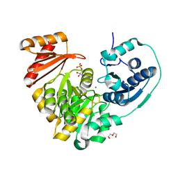
6NN2
 | | Xanthomonas citri PGM Apo-Phospho | | Descriptor: | CALCIUM ION, HEXAETHYLENE GLYCOL, Phosphoglucomutase | | Authors: | Stiers, K.M, Beamer, L.J. | | Deposit date: | 2019-01-14 | | Release date: | 2019-04-10 | | Last modified: | 2023-10-11 | | Method: | X-RAY DIFFRACTION (1.44 Å) | | Cite: | Structural and dynamical description of the enzymatic reaction of a phosphohexomutase.
Struct Dyn., 6, 2019
|
|
6NNT
 | |
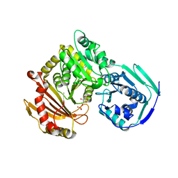
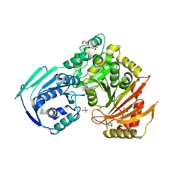
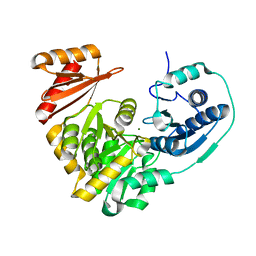
6NN1
 | | Xanthomonas citri PGM Apo-Dephospho | | Descriptor: | DI(HYDROXYETHYL)ETHER, MAGNESIUM ION, PHOSPHATE ION, ... | | Authors: | Stiers, K.M, Beamer, L.J. | | Deposit date: | 2019-01-14 | | Release date: | 2019-04-10 | | Last modified: | 2023-10-11 | | Method: | X-RAY DIFFRACTION (1.5 Å) | | Cite: | Structural and dynamical description of the enzymatic reaction of a phosphohexomutase.
Struct Dyn., 6, 2019
|
|
6NQF
 | |
6NQE
 | |
6NOQ
 | |
6NP8
 | |
6NNP
 | |
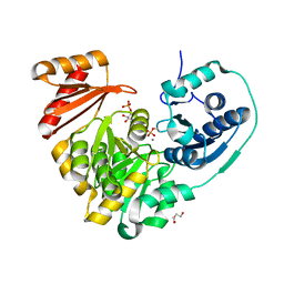
6NNU
 | | Xanthomonas citri Phospho-PGM in complex with glucose-1,6-phosphate | | Descriptor: | 1,6-di-O-phosphono-alpha-D-glucopyranose, CALCIUM ION, DI(HYDROXYETHYL)ETHER, ... | | Authors: | Stiers, K.M, Beamer, L.J. | | Deposit date: | 2019-01-15 | | Release date: | 2019-04-10 | | Last modified: | 2023-10-11 | | Method: | X-RAY DIFFRACTION (1.46 Å) | | Cite: | Structural and dynamical description of the enzymatic reaction of a phosphohexomutase.
Struct Dyn., 6, 2019
|
|
6NQH
 | |
6NNS
 | |
6NPX
 | |
6NQG
 | |
6NNN
 | |
6NNO
 | |
5JN5
 | |


