4YGH
 
 | |
6PQB
 | |
7JLZ
 | |
4YH0
 | |
4YDA
 | |
4YDB
 | |
4YFQ
 | |
4YKO
 | |
4YBP
 | |
4YGZ
 | |
4YGG
 | |
4YCE
 | |
4YFR
 | |
4YKM
 | |
4YBU
 | |
4YCH
 | |
4YFP
 | |
4M6S
 | |
4M7M
 | |
4I9S
 | |
4I9R
 | |
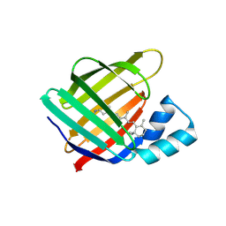
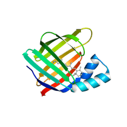
6C7Z
 | | Crystal structure of the Q108K:K40L:T51V:R58F mutant of human Cellular Retinol Binding Protein II in complex with synthetic Ligand Julolidine | | Descriptor: | (2E,4E)-3-methyl-5-(2,3,6,7-tetrahydro-1H,5H-pyrido[3,2,1-ij]quinolin-9-yl)penta-2,4-dienal, ACETATE ION, Retinol-binding protein 2 | | Authors: | Nosrati, M, Geiger, J.H. | | Deposit date: | 2018-01-23 | | Release date: | 2018-04-25 | | Last modified: | 2023-10-04 | | Method: | X-RAY DIFFRACTION (1.42 Å) | | Cite: | A Genetically Encoded Ratiometric pH Probe: Wavelength Regulation-Inspired Design of pH Indicators.
Chembiochem, 19, 2018
|
|
6E5S
 | |
5F58
 | |
5F6B
 | |


