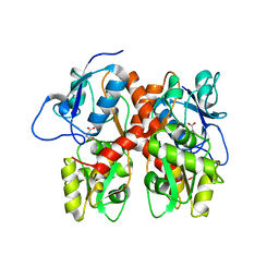
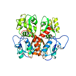
Loading
PDBj
MenuPDBj@FacebookPDBj@TwitterPDBj@YouTubewwPDB FoundationwwPDB
RCSB PDBPDBeBMRBAdv. SearchSearch help
Search by PDB author
5ICT
DownloadVisualize
BU of 5ict by Molmil
Crystal structure of the Drosophila GluR1A ligand binding domain Y792T mutant complex with glutamate
Descriptor: GLUTAMIC ACID, GLYCEROL, Glutamate receptor 1
Authors:Dharkar, P, Mayer, M.L.
Deposit date:2016-02-23
Release date:2016-12-21
Last modified:2023-09-27
Method:X-RAY DIFFRACTION (1.68 Å)
Cite:Novel Functional Properties of Drosophila CNS Glutamate Receptors.
Neuron, 92, 2016
4WXJ
DownloadVisualize
BU of 4wxj by Molmil
Drosophila muscle GluRIIB complex with glutamate
Descriptor: GLUTAMIC ACID, Glutamate receptor IIB,Glutamate receptor IIB
Authors:Dharkar, P, Mayer, M.L.
Deposit date:2014-11-13
Release date:2015-04-29
Last modified:2023-09-27
Method:X-RAY DIFFRACTION (2.002 Å)
Cite:Functional reconstitution of Drosophila melanogaster NMJ glutamate receptors.
Proc.Natl.Acad.Sci.USA, 112, 2015
5DT6
DownloadVisualize
BU of 5dt6 by Molmil
Crystal structure of the Drosophila GluR1A ligand binding domain complex with glutamate
Descriptor: GLUTAMIC ACID, GLYCEROL, Glutamate receptor 1
Authors:Dharkar, P, Mayer, M.L.
Deposit date:2015-09-17
Release date:2016-09-28
Last modified:2023-09-27
Method:X-RAY DIFFRACTION (1.598 Å)
Cite:Novel Functional Properties of Drosophila CNS Glutamate Receptors.
Neuron, 92, 2016
5DTB
DownloadVisualize
BU of 5dtb by Molmil
Crystal structure of the Drosophila CG3822 KaiR1D ligand binding domain complex with glutamate
Descriptor: CG3822, GLUTAMIC ACID, GLYCEROL, ...
Authors:Dharkar, P, Mayer, M.L.
Deposit date:2015-09-17
Release date:2016-09-28
Last modified:2023-09-27
Method:X-RAY DIFFRACTION (1.843 Å)
Cite:Novel Functional Properties of Drosophila CNS Glutamate Receptors.
Neuron, 92, 2016
5EHM
DownloadVisualize
BU of 5ehm by Molmil
Crystal structure of the Drosophila CG3822 KaiR1D ligand binding domain complex with NMDA
Descriptor: N-methyl-D-aspartic acid, RE06730p,CG3822
Authors:Dharkar, P, Mayer, M.L.
Deposit date:2015-10-28
Release date:2016-11-09
Last modified:2023-11-15
Method:X-RAY DIFFRACTION (1.281 Å)
Cite:Novel Functional Properties of Drosophila CNS Glutamate Receptors.
Neuron, 92, 2016
5EHS
DownloadVisualize
BU of 5ehs by Molmil
Crystal structure of the Drosophila CG3822 KaiR1D ligand binding domain complex with D-AP5
Descriptor: 5-phosphono-D-norvaline, 5-phosphono-L-norvaline, RE06730p,GH17276
Authors:Dharkar, P, Mayer, M.L.
Deposit date:2015-10-28
Release date:2016-11-09
Last modified:2023-09-27
Method:X-RAY DIFFRACTION (1.749 Å)
Cite:Novel Functional Properties of Drosophila CNS Glutamate Receptors.
Neuron, 92, 2016
5E0S
DownloadVisualize
BU of 5e0s by Molmil
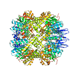
crystal structure of the active form of the proteolytic complex clpP1 and clpP2
Descriptor: ATP-dependent Clp protease proteolytic subunit 1, ATP-dependent Clp protease proteolytic subunit 2
Authors:LI, M, Wlodawer, A, Maurizi, M.
Deposit date:2015-09-29
Release date:2016-02-17
Last modified:2016-04-13
Method:X-RAY DIFFRACTION (2.9 Å)
Cite:Structure and Functional Properties of the Active Form of the Proteolytic Complex, ClpP1P2, from Mycobacterium tuberculosis.
J.Biol.Chem., 291, 2016
5DZK
DownloadVisualize
BU of 5dzk by Molmil
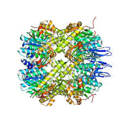
Crystal structure of the active form of the proteolytic complex clpP1 and clpP2
Descriptor: ATP-dependent Clp protease proteolytic subunit 1, ATP-dependent Clp protease proteolytic subunit 2, BEZ-LEU-LEU
Authors:LI, M, Wlodawer, A, Maurizi, M.
Deposit date:2015-09-25
Release date:2016-02-17
Last modified:2016-04-13
Method:X-RAY DIFFRACTION (3.07 Å)
Cite:Structure and Functional Properties of the Active Form of the Proteolytic Complex, ClpP1P2, from Mycobacterium tuberculosis.
J.Biol.Chem., 291, 2016

218853

PDB entries from 2024-04-24

PDB statisticsPDBj update infoContact PDBjnumon