1AJ3
 
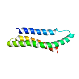 | | SOLUTION STRUCTURE OF THE SPECTRIN REPEAT, NMR, 20 STRUCTURES | | Descriptor: | ALPHA SPECTRIN | | Authors: | Pascual, J, Pfuhl, M, Walther, D, Saraste, M, Nilges, M. | | Deposit date: | 1997-05-14 | | Release date: | 1997-07-07 | | Last modified: | 2022-02-16 | | Method: | SOLUTION NMR | | Cite: | Solution structure of the spectrin repeat: a left-handed antiparallel triple-helical coiled-coil.
J.Mol.Biol., 273, 1997
|
|
1JE5
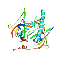 | | Crystal Structure of gp2.5, a Single-Stranded DNA Binding Protein Encoded by Bacteriophage T7 | | Descriptor: | CALCIUM ION, HELIX-DESTABILIZING PROTEIN | | Authors: | Hollis, T, Stattel, J.M, Walther, D.S, Richardson, C.C, Ellenberger, T.E. | | Deposit date: | 2001-06-15 | | Release date: | 2001-08-22 | | Last modified: | 2024-02-07 | | Method: | X-RAY DIFFRACTION (1.9 Å) | | Cite: | Structure of the gene 2.5 protein, a single-stranded DNA binding protein encoded by bacteriophage T7.
Proc.Natl.Acad.Sci.USA, 98, 2001
|
|


