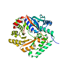
Loading
PDBj
MenuPDBj@FacebookPDBj@TwitterPDBj@YouTubewwPDB FoundationwwPDB
RCSB PDBPDBeBMRBAdv. SearchSearch help
PDB: 491 results

2IMG
DownloadVisualize
BU of 2img by Molmil
Crystal structure of dual specificity protein phosphatase 23 from Homo sapiens in complex with ligand malate ion
Descriptor: D-MALATE, Dual specificity protein phosphatase 23
Authors:Agarwal, R, Burley, S.K, Swaminathan, S, New York SGX Research Center for Structural Genomics (NYSGXRC)
Deposit date:2006-10-04
Release date:2006-10-17
Last modified:2021-02-03
Method:X-RAY DIFFRACTION (1.93 Å)
Cite:Structure of human dual specificity protein phosphatase 23, VHZ, enzyme-substrate/product complex.
J.Biol.Chem., 283, 2008
2ICS
DownloadVisualize
BU of 2ics by Molmil
Crystal structure of an adenine deaminase
Descriptor: ADENINE, Adenine Deaminase, ZINC ION
Authors:Sugadev, R, Kumaran, D, Burley, S.K, Swaminathan, S, New York SGX Research Center for Structural Genomics (NYSGXRC)
Deposit date:2006-09-13
Release date:2006-10-17
Last modified:2021-02-03
Method:X-RAY DIFFRACTION (2.3 Å)
Cite:Crystal structure of an adenine deaminase
TO BE PUBLISHED
2IOJ
DownloadVisualize
BU of 2ioj by Molmil
Crystal structure of protein AF1212 from Archaeoglobus fulgidus, Pfam DRTGG
Descriptor: Hypothetical protein AF_1212
Authors:Tyagi, R, Burley, S.K, Swaminathan, S, New York SGX Research Center for Structural Genomics (NYSGXRC)
Deposit date:2006-10-10
Release date:2006-10-31
Last modified:2021-02-03
Method:X-RAY DIFFRACTION (2.15 Å)
Cite:The crystal structure of a hypothetical protein (Y1212_ARCFU) from Archeoglobus fulgidus
To be Published
1RRM
DownloadVisualize
BU of 1rrm by Molmil
Crystal Structure of Lactaldehyde reductase
Descriptor: ADENOSINE-5-DIPHOSPHORIBOSE, FE (II) ION, Lactaldehyde reductase, ...
Authors:Kumaran, D, Swaminathan, S, Burley, S.K, New York SGX Research Center for Structural Genomics (NYSGXRC)
Deposit date:2003-12-08
Release date:2004-08-17
Last modified:2021-02-03
Method:X-RAY DIFFRACTION (1.6 Å)
Cite:Crystal structure of Lactaldehyde reductase
To be Published
1RTT
DownloadVisualize
BU of 1rtt by Molmil
Crystal structure determination of a putative NADH-dependent reductase using sulfur anomalous signal
Descriptor: SULFATE ION, conserved hypothetical protein
Authors:Agarwal, R, Swaminathan, S, Burley, S.K, New York SGX Research Center for Structural Genomics (NYSGXRC)
Deposit date:2003-12-10
Release date:2004-08-17
Last modified:2024-02-14
Method:X-RAY DIFFRACTION (1.28 Å)
Cite:Structure determination of an FMN reductase from Pseudomonas aeruginosa PA01 using sulfur anomalous signal.
ACTA CRYSTALLOGR.,SECT.D, 62, 2006
1SGJ
DownloadVisualize
BU of 1sgj by Molmil
Crystal structure of citrate lyase beta subunit
Descriptor: MAGNESIUM ION, OXALOACETATE ION, citrate lyase, ...
Authors:Eswaramoorthy, S, Swaminathan, S, Burley, S.K, New York SGX Research Center for Structural Genomics (NYSGXRC)
Deposit date:2004-02-23
Release date:2004-08-17
Last modified:2024-02-14
Method:X-RAY DIFFRACTION (1.84 Å)
Cite:Crystal structure of citrate lyase beta subunit
To be Published
1YXW
DownloadVisualize
BU of 1yxw by Molmil
A common binding site for disialyllactose and a tri-peptide in the C-fragment of tetanus neurotoxin
Descriptor: GLUTAMIC ACID, TRYPTOPHAN, TYROSINE, ...
Authors:Jayaraman, S, Eswaramoorthy, S, Kumaran, D, Swaminathan, S.
Deposit date:2005-02-22
Release date:2005-03-15
Last modified:2023-08-23
Method:X-RAY DIFFRACTION (2.2 Å)
Cite:Common binding site for disialyllactose and tri-peptide in C-fragment of tetanus neurotoxin
Proteins, 61, 2005
1ZCC
DownloadVisualize
BU of 1zcc by Molmil
Crystal structure of glycerophosphodiester phosphodiesterase from Agrobacterium tumefaciens str.C58
Descriptor: ACETATE ION, SULFATE ION, glycerophosphodiester phosphodiesterase
Authors:Krishnamurthy, N.R, Kumaran, D, Swaminathan, S, Burley, S.K, New York SGX Research Center for Structural Genomics (NYSGXRC)
Deposit date:2005-04-11
Release date:2005-05-03
Last modified:2024-02-14
Method:X-RAY DIFFRACTION (2.5 Å)
Cite:Crystal structure of glycerophosphodiester phosphodiesterase from Agrobacterium tumefaciens by SAD with a large asymmetric unit.
Proteins, 65, 2006
1YYN
DownloadVisualize
BU of 1yyn by Molmil
A common binding site for disialyllactose and a tri-peptide in the C-fragment of tetanus neurotoxin
Descriptor: N-acetyl-alpha-neuraminic acid-(2-8)-N-acetyl-alpha-neuraminic acid-(2-3)-alpha-D-galactopyranose-(1-4)-beta-D-glucopyranose, Tetanus toxin
Authors:Seetharaman, J, Eswaramoorthy, S, Kumaran, D, Swaminathan, S.
Deposit date:2005-02-25
Release date:2005-03-15
Last modified:2023-10-25
Method:X-RAY DIFFRACTION (2.3 Å)
Cite:Common binding site for disialyllactose and tri-peptide in C-fragment of tetanus neurotoxin
Proteins, 61, 2005
1TXL
DownloadVisualize
BU of 1txl by Molmil
Crystal structure of metal-binding protein yodA from E. coli, Pfam DUF149
Descriptor: Metal-binding protein yodA, ZINC ION
Authors:Eswaramoorthy, S, Swaminathan, S, Burley, S.K, New York SGX Research Center for Structural Genomics (NYSGXRC)
Deposit date:2004-07-05
Release date:2004-07-20
Last modified:2021-02-03
Method:X-RAY DIFFRACTION (1.7 Å)
Cite:Crystal structure of a hypothetical protein yodA
To be Published
2A9F
DownloadVisualize
BU of 2a9f by Molmil
Crystal structure of a putative malic enzyme ((S)-malate:NAD+ oxidoreductase (decarboxylating))
Descriptor: MAGNESIUM ION, putative malic enzyme ((S)-malate:NAD+ oxidoreductase (decarboxylating))
Authors:Seetharaman, J, Swaminathan, S, Burley, S.K, New York SGX Research Center for Structural Genomics (NYSGXRC)
Deposit date:2005-07-11
Release date:2005-08-02
Last modified:2024-02-14
Method:X-RAY DIFFRACTION (2.5 Å)
Cite:Crystal structure of a putative malic enzyme ((S)-malate:NAD+ oxidoreductase (decarboxylating))
To be Published
1TYH
DownloadVisualize
BU of 1tyh by Molmil
Crystal Structure of Transcriptional Activator tenA from Bacillus subtilis
Descriptor: Transcriptional activator tenA
Authors:Eswaramoorthy, S, Swaminathan, S, Burley, S.K, New York SGX Research Center for Structural Genomics (NYSGXRC)
Deposit date:2004-07-07
Release date:2004-07-20
Last modified:2021-02-03
Method:X-RAY DIFFRACTION (2.54 Å)
Cite:Crystal Structure of Transcriptional Activator tenA from Bacillus subtilis
To be Published
3T8L
DownloadVisualize
BU of 3t8l by Molmil
Crystal Structure of adenine deaminase with Mn/Fe
Descriptor: Adenine deaminase 2, UNKNOWN ATOM OR ION
Authors:Bagaria, A, Kumaran, D, Burley, S.K, Swaminathan, S, New York SGX Research Center for Structural Genomics (NYSGXRC)
Deposit date:2011-08-01
Release date:2011-11-02
Last modified:2023-12-06
Method:X-RAY DIFFRACTION (2.8 Å)
Cite:The catalase activity of diiron adenine deaminase.
Protein Sci., 20, 2011
2NRQ
DownloadVisualize
BU of 2nrq by Molmil
Crystal structure of protein SSO0741 from Sulfolobus solfataricus, Pfam DUF54
Descriptor: Hypothetical protein ORF-c20_032
Authors:Rao, K.N, Burley, S.K, Swaminathan, S, New York SGX Research Center for Structural Genomics (NYSGXRC)
Deposit date:2006-11-02
Release date:2006-11-28
Last modified:2023-12-27
Method:X-RAY DIFFRACTION (2.6 Å)
Cite:UPF201 archaeal specific family members reveal structural similarity to RNA-binding proteins but low likelihood for RNA-binding function.
Plos One, 3, 2008
2OG9
DownloadVisualize
BU of 2og9 by Molmil
Crystal Structure of mandelate racemase/muconate lactonizing enzyme from Polaromonas sp. JS666
Descriptor: CALCIUM ION, Mandelate racemase/muconate lactonizing enzyme
Authors:Kumaran, D, Burley, S.K, Swaminathan, S, New York SGX Research Center for Structural Genomics (NYSGXRC)
Deposit date:2007-01-05
Release date:2007-02-27
Last modified:2023-12-27
Method:X-RAY DIFFRACTION (1.9 Å)
Cite:Crystal Structure of mandelate racemase/muconate lactonizing enzyme from Polaromonas sp. JS666
To be Published
2OGK
DownloadVisualize
BU of 2ogk by Molmil
Crystal structure of protein AF2318 from Archaeglobus fulgidus, Pfam DUF54
Descriptor: Hypothetical protein
Authors:Rao, K.N, Burley, S.K, Swaminathan, S, New York SGX Research Center for Structural Genomics (NYSGXRC)
Deposit date:2007-01-05
Release date:2007-01-30
Last modified:2023-12-27
Method:X-RAY DIFFRACTION (3 Å)
Cite:UPF201 archaeal specific family members reveal structural similarity to RNA-binding proteins but low likelihood for RNA-binding function.
Plos One, 3, 2008
2NWU
DownloadVisualize
BU of 2nwu by Molmil
Crystal structure of protein SSO1042 from Sulfolobus solfataricus, Pfam DUF54
Descriptor: UPF0201 protein SSO1042
Authors:Rao, K.N, Burley, S.K, Swaminathan, S, New York SGX Research Center for Structural Genomics (NYSGXRC)
Deposit date:2006-11-16
Release date:2006-12-12
Last modified:2023-12-27
Method:X-RAY DIFFRACTION (2.4 Å)
Cite:UPF201 archaeal specific family members reveal structural similarity to RNA-binding proteins but low likelihood for RNA-binding function.
Plos One, 3, 2008
2OOD
DownloadVisualize
BU of 2ood by Molmil
Crystal structure of guanine deaminase from Bradyrhizobium japonicum
Descriptor: Blr3880 protein, GUANINE, ZINC ION
Authors:Agarwal, R, Burley, S.K, Swaminathan, S, New York SGX Research Center for Structural Genomics (NYSGXRC)
Deposit date:2007-01-25
Release date:2007-02-20
Last modified:2023-12-27
Method:X-RAY DIFFRACTION (2.62 Å)
Cite:Crystal structure of guanine deaminase from Bradyrhizobium japonicum
To be Published
2OPJ
DownloadVisualize
BU of 2opj by Molmil
Crystal structure of O-succinylbenzoate synthase
Descriptor: O-succinylbenzoate-CoA synthase
Authors:Sugadev, R, Burley, S.K, Swaminathan, S, New York SGX Research Center for Structural Genomics (NYSGXRC)
Deposit date:2007-01-29
Release date:2007-02-20
Last modified:2023-12-27
Method:X-RAY DIFFRACTION (1.6 Å)
Cite:Crystal structure of O-succinylbenzoate synthase
To be Published
1VQV
DownloadVisualize
BU of 1vqv by Molmil
Crystal Structure of Thiamine Monophosphate Kinase (thil) from Aquifex Aeolicus
Descriptor: PHOSPHATE ION, thiamine monophosphate kinase
Authors:Eswaramoorthy, S, Swaminathan, S, Burley, S.K, New York SGX Research Center for Structural Genomics (NYSGXRC)
Deposit date:2005-01-05
Release date:2005-01-11
Last modified:2023-12-27
Method:X-RAY DIFFRACTION (2.65 Å)
Cite:Crystal Structure of Thiamine Monophosphate Kinase (Thil) from Aquifex Aeolicus
To be Published
1X77
DownloadVisualize
BU of 1x77 by Molmil
Crystal structure of a NAD(P)H-dependent FMN reductase complexed with FMN
Descriptor: FLAVIN MONONUCLEOTIDE, conserved hypothetical protein
Authors:Agarwal, R, Swaminathan, S, Burley, S.K, New York SGX Research Center for Structural Genomics (NYSGXRC)
Deposit date:2004-08-13
Release date:2004-08-24
Last modified:2023-08-23
Method:X-RAY DIFFRACTION (2.7 Å)
Cite:Structure determination of an FMN reductase from Pseudomonas aeruginosa PA01 using sulfur anomalous signal.
ACTA CRYSTALLOGR.,SECT.D, 62, 2006
1YIR
DownloadVisualize
BU of 1yir by Molmil
Crystal Structure of a Nicotinate Phosphoribosyltransferase
Descriptor: Nicotinate phosphoribosyltransferase 2, SULFATE ION
Authors:Seetharaman, J, Swaminathan, S, Burley, S.K, New York SGX Research Center for Structural Genomics (NYSGXRC)
Deposit date:2005-01-12
Release date:2005-02-01
Last modified:2024-02-14
Method:X-RAY DIFFRACTION (2.1 Å)
Cite:Crystal Structure of a Nicotinate Phosphoribosyltransferase
To be Published
1SGM
DownloadVisualize
BU of 1sgm by Molmil
Crystal Structure of Hypothetical Protein YXAF
Descriptor: Putative HTH-type transcriptional regulator yxaF
Authors:Seetharaman, J, Swaminathan, S, Burley, S.K, New York SGX Research Center for Structural Genomics (NYSGXRC)
Deposit date:2004-02-24
Release date:2004-08-17
Last modified:2024-02-14
Method:X-RAY DIFFRACTION (2 Å)
Cite:Crystal structure of a putative HTH-type transcriptional regulator yxaF from Bacillus subtilis.
Proteins, 63, 2006
1T3A
DownloadVisualize
BU of 1t3a by Molmil
Crystal structure of Clostridium botulinum neurotoxin type E catalytic domain
Descriptor: CHLORIDE ION, ZINC ION, neurotoxin type E
Authors:Agarwal, R, Eswaramoorthy, S, Kumaran, D, Binz, T, Swaminathan, S.
Deposit date:2004-04-26
Release date:2004-06-29
Last modified:2024-02-14
Method:X-RAY DIFFRACTION (2.16 Å)
Cite:Structural analysis of botulinum neurotoxin type E catalytic domain and its mutant Glu212-->Gln reveals the pivotal role of the Glu212 carboxylate in the catalytic pathway
Biochemistry, 43, 2004
1T3C
DownloadVisualize
BU of 1t3c by Molmil
Clostridium botulinum type E catalytic domain E212Q mutant
Descriptor: CHLORIDE ION, ZINC ION, neurotoxin type E
Authors:Agarwal, R, Eswaramoorthy, S, Kumaran, D, Binz, T, Swaminathan, S.
Deposit date:2004-04-26
Release date:2004-06-29
Last modified:2023-08-23
Method:X-RAY DIFFRACTION (1.9 Å)
Cite:Structural analysis of botulinum neurotoxin type E catalytic domain and its mutant Glu212-->Gln reveals the pivotal role of the Glu212 carboxylate in the catalytic pathway
Biochemistry, 43, 2004

219869

PDB entries from 2024-05-15

PDB statisticsPDBj update infoContact PDBjnumon