Loading
PDBj
MenuPDBj@FacebookPDBj@TwitterPDBj@YouTubewwPDB FoundationwwPDB
RCSB PDBPDBeBMRBAdv. SearchSearch help
PDB: 51 results

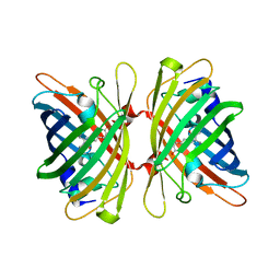
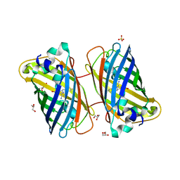
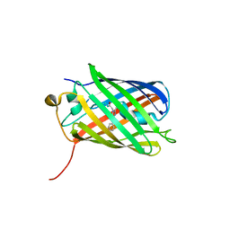
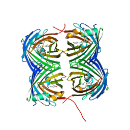
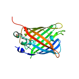
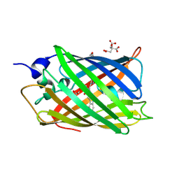
4N3D
DownloadVisualize
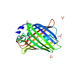
BU of 4n3d by Molmil
Crystal structure of the dimeric variant EGFP-K162Q in P61 space group
Descriptor: GLYCEROL, Green fluorescent protein, PHOSPHATE ION, ...
Authors:Pletneva, N.V, Pletnev, V.Z, Pletnev, S.V.
Deposit date:2013-10-07
Release date:2014-08-27
Method:X-RAY DIFFRACTION (1.34 Å)
Cite:Three dimensional structure of the dimeric gene-engineered variant of green fluorescent protein egfp-K162Q in P61 crystal space group
Rus.J.Bioorg.Chem., 40, 2014
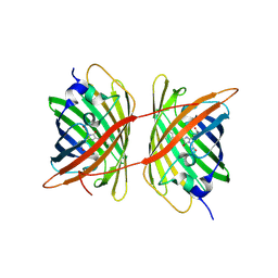
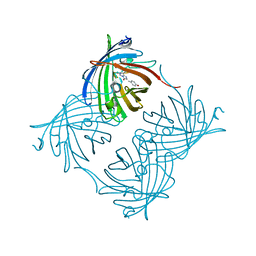
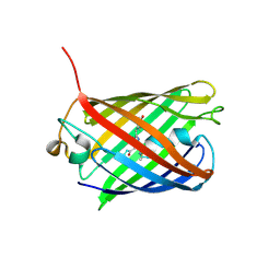
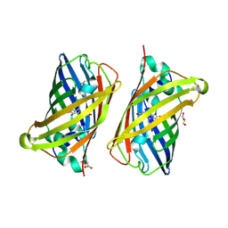
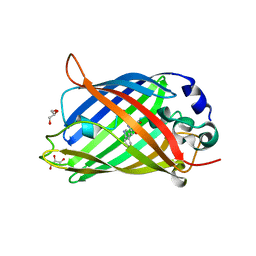
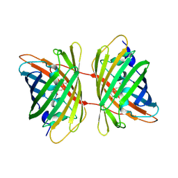
4HE4
DownloadVisualize
BU of 4he4 by Molmil
Crystal structure of the yellow fluorescent protein phiYFP (Phialidium sp.)
Descriptor: Yellow fluorescent protein
Authors:Pletneva, N, Pletnev, S, Pletnev, V.Z.
Deposit date:2012-10-03
Release date:2013-05-29
Last modified:2023-12-06
Method:X-RAY DIFFRACTION (2.05 Å)
Cite:Yellow fluorescent protein phiYFPv (Phialidium): structure and structure-based mutagenesis.
Acta Crystallogr.,Sect.D, 69, 2013
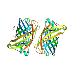
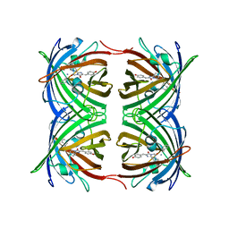
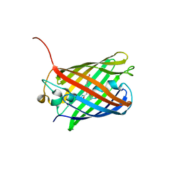
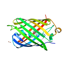
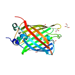
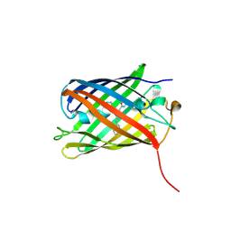
4ZFS
DownloadVisualize
BU of 4zfs by Molmil
Phototoxic Fluorescent Protein KillerOrange
Descriptor: KillerOrange
Authors:Pletneva, N.V, Pletnev, V.Z, Pletnev, S.
Deposit date:2015-04-21
Release date:2015-12-23
Last modified:2024-05-15
Method:X-RAY DIFFRACTION (2.01 Å)
Cite:Crystal Structure of Phototoxic Orange Fluorescent Proteins with a Tryptophan-Based Chromophore.
Plos One, 10, 2015
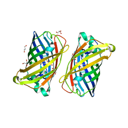
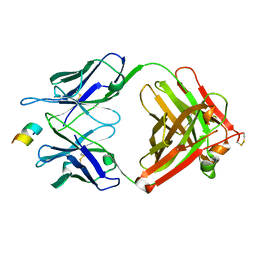
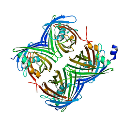
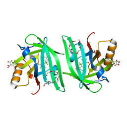
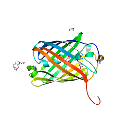
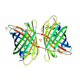
3LVD
DownloadVisualize
BU of 3lvd by Molmil
Crystal structure of GFP-like protein aceGFP_G222E (A. coerulescens). UV-photoconverted green form.
Descriptor: GLYCEROL, Green fluorescent protein
Authors:Pletneva, N.V, Pletnev, V.Z, Pletnev, S.V.
Deposit date:2010-02-19
Release date:2010-03-09
Last modified:2023-11-15
Method:X-RAY DIFFRACTION (1.751 Å)
Cite:Structural evidence for a dehydrated intermediate in green fluorescent protein chromophore biosynthesis.
J.Biol.Chem., 285, 2010
3LVC
DownloadVisualize
BU of 3lvc by Molmil
Crystal structure of GFP-like protein aceGFP_G222E (A. coerulescens). Colorless form.
Descriptor: GLYCEROL, Green fluorescent protein
Authors:Pletneva, N.V, Pletnev, V.Z, Pletnev, S.V.
Deposit date:2010-02-19
Release date:2010-03-09
Last modified:2023-11-15
Method:X-RAY DIFFRACTION (1.14 Å)
Cite:Structural evidence for a dehydrated intermediate in green fluorescent protein chromophore biosynthesis.
J.Biol.Chem., 285, 2010
3LVA
DownloadVisualize
BU of 3lva by Molmil
Crystal structure of colorless GFP-like protein from Aequorea coerulescens
Descriptor: GLYCEROL, Green fluorescent protein, SULFATE ION
Authors:Pletneva, N.V, Pletnev, V.Z, Pletnev, S.V.
Deposit date:2010-02-19
Release date:2010-03-09
Last modified:2023-11-15
Method:X-RAY DIFFRACTION (1.5 Å)
Cite:Structural evidence for a dehydrated intermediate in green fluorescent protein chromophore biosynthesis.
J.Biol.Chem., 285, 2010
2OJK
DownloadVisualize
BU of 2ojk by Molmil
Crystal Structure of Green Fluorescent Protein from Zoanthus sp at 2.2 A Resolution
Descriptor: GFP-like fluorescent chromoprotein FP506
Authors:Pletneva, N.V, Pletnev, S.V, Tikhonova, T.V, Pletnev, V.Z.
Deposit date:2007-01-12
Release date:2007-09-25
Last modified:2023-11-15
Method:X-RAY DIFFRACTION (2.2 Å)
Cite:Refined crystal structures of red and green fluorescent proteins from the button polyp Zoanthus.
Acta Crystallogr.,Sect.D, 63, 2007
2OGR
DownloadVisualize
BU of 2ogr by Molmil
Crystal Structure of Yellow Fluorescent Protein from Zoanthus sp. at 1.8 A Resolution
Descriptor: FLUORESCENT PROTEIN FP538
Authors:Pletneva, N.V, Pletnev, S.V, Tikhonova, T.V, Pletnev, V.Z.
Deposit date:2007-01-08
Release date:2007-09-25
Last modified:2023-12-27
Method:X-RAY DIFFRACTION (1.8 Å)
Cite:Three-dimensional structure of yellow fluorescent protein zYFP538 from Zoanthus sp. at the resolution 1.8 angstrom
Bioorg.Khim., 33
7YZJ
DownloadVisualize
BU of 7yzj by Molmil
FAB IN COMPLEX WITH ANTIGENIC PEPTIDE OF INTERLEUKIN-2
Descriptor: Antigenic peptide, Heavy chain of FAB fragment, Light chain of FAB fragment
Authors:Pletnev, V, Pletneva, N.
Deposit date:2022-02-20
Release date:2023-02-01
Last modified:2024-02-07
Method:X-RAY DIFFRACTION (2.6 Å)
Cite:Three-Dimensional Structure of Fab Fragment of Monoclonal Antibody LNKB-2 Complexed with Antigenic Nonapeptide from Human Interleukin-2
Russ.J.Bioorganic Chem., 2023
3BXA
DownloadVisualize
BU of 3bxa by Molmil
Monomeric Far-red Fluorescent Protein mKate Crystallized at pH 4.2
Descriptor: CITRIC ACID, Far-red fluorescent protein mKate
Authors:Pletnev, S, Pletneva, N, Pletnev, V.
Deposit date:2008-01-12
Release date:2008-07-22
Last modified:2017-10-25
Method:X-RAY DIFFRACTION (1.75 Å)
Cite:A Crystallographic Study of Bright Far-Red Fluorescent Protein mKate Reveals pH-induced cis-trans Isomerization of the Chromophore.
J.Biol.Chem., 283, 2008
3BXC
DownloadVisualize
BU of 3bxc by Molmil
Monomeric Far-red Fluorescent Protein mKate Crystallized at pH 9.0
Descriptor: Far-red fluorescent protein mKate
Authors:Pletnev, S, Pletneva, N, Pletnev, V.
Deposit date:2008-01-12
Release date:2008-07-22
Last modified:2019-07-24
Method:X-RAY DIFFRACTION (2.6 Å)
Cite:A Crystallographic Study of Bright Far-Red Fluorescent Protein mKate Reveals pH-induced cis-trans Isomerization of the Chromophore.
J.Biol.Chem., 283, 2008
3BXB
DownloadVisualize
BU of 3bxb by Molmil
Monomeric Far-red Fluorescent Protein mKate Crystallized at pH 7.0
Descriptor: Far-red fluorescent protein mKate
Authors:Pletnev, S, Pletneva, N, Pletnev, V.
Deposit date:2008-01-12
Release date:2008-07-22
Last modified:2019-07-24
Method:X-RAY DIFFRACTION (2.6 Å)
Cite:A Crystallographic Study of Bright Far-Red Fluorescent Protein mKate Reveals pH-induced cis-trans Isomerization of the Chromophore.
J.Biol.Chem., 283, 2008
7SF9
DownloadVisualize
BU of 7sf9 by Molmil
Branchiostoma Floridae Violet Fluorescent Protein
Descriptor: Violet Fluorescent Protein
Authors:Pletnev, S, Pletneva, N, Pletnev, V.Z, Muslinkina, L.
Deposit date:2021-10-03
Release date:2022-03-30
Last modified:2023-10-18
Method:X-RAY DIFFRACTION (1.8 Å)
Cite:A novel violet fluorescent protein contains a unique oxidized tyrosine as the simplest chromophore ever reported in fluorescent proteins.
Protein Sci., 31, 2022
7SFA
DownloadVisualize
BU of 7sfa by Molmil
Branchiostoma floridae fluorescent protein LanFP10A2
Descriptor: Fluorescent protein lanFP10A2
Authors:Pletnev, S, Pletneva, N, Pletnev, V.Z, Muslinkina, L.
Deposit date:2021-10-03
Release date:2022-03-30
Last modified:2023-11-15
Method:X-RAY DIFFRACTION (1.65 Å)
Cite:A novel violet fluorescent protein contains a unique oxidized tyrosine as the simplest chromophore ever reported in fluorescent proteins.
Protein Sci., 31, 2022
8BVG
DownloadVisualize
BU of 8bvg by Molmil
Bright fluorescent protein BrUSLEE with subnanosecond fluorescence lifetime
Descriptor: BrUSSLEE, GLYCEROL
Authors:Pletnev, V, Pletneva, N.
Deposit date:2022-12-03
Release date:2023-04-12
Last modified:2023-04-26
Method:X-RAY DIFFRACTION (2.38 Å)
Cite:Crystal Structure of Bright Fluorescent Protein BrUSLEE with Subnanosecond Fluorescence Lifetime; Electric and Dynamic Properties.
Int J Mol Sci, 24, 2023
6U1A
DownloadVisualize
BU of 6u1a by Molmil
Crystal Structure of Fluorescent Protein FusionRed
Descriptor: CALCIUM ION, NICKEL (II) ION, Red fluorescent protein
Authors:Pletnev, S, Muslinkina, L, Pletneva, N, Pletnev, V.Z.
Deposit date:2019-08-15
Release date:2020-04-22
Last modified:2023-11-15
Method:X-RAY DIFFRACTION (1.09 Å)
Cite:Two independent routes of post-translational chemistry in fluorescent protein FusionRed.
Int.J.Biol.Macromol., 155, 2020
6UBO
DownloadVisualize
BU of 6ubo by Molmil
Fluorogen Activating Protein Dib1
Descriptor: 12-(diethylamino)-2,2-bis(fluoranyl)-4,5-dimethyl-5-aza-3-azonia-2-boranuidatricyclo[7.4.0.0^{3,7}]trideca-1(13),3,7,9,11-pentaen-6-one, CITRIC ACID, Outer membrane lipoprotein Blc, ...
Authors:Muslinkina, L, Pletneva, N, Pletnev, V.Z, Pletnev, S.
Deposit date:2019-09-12
Release date:2020-09-16
Last modified:2023-10-11
Method:X-RAY DIFFRACTION (1.58 Å)
Cite:Structure-Based Rational Design of Two Enhanced Bacterial Lipocalin Blc Tags for Protein-PAINT Super-resolution Microscopy.
Acs Chem.Biol., 15, 2020
6ZQO
DownloadVisualize
BU of 6zqo by Molmil
EYFP mutant - F165G
Descriptor: G protein/GFP fusion protein, SULFATE ION
Authors:Pletnev, V.Z, Pletnev, S.V, Pletneva, N.V.
Deposit date:2020-07-10
Release date:2021-06-16
Last modified:2024-01-31
Method:X-RAY DIFFRACTION (2.2 Å)
Cite:Amino acid residue at the 165th position tunes EYFP chromophore maturation. A structure-based design.
Comput Struct Biotechnol J, 19, 2021
4RYW
DownloadVisualize
BU of 4ryw by Molmil
Crystal structure of the photoconverted green fluorescent protein NowGFP_conv (the variant of cyan Cerulean) at pH 7.0
Descriptor: GLYCEROL, NowGFP_conv
Authors:Pletnev, V.Z, Pletneva, N.V, Pletnev, S.V.
Deposit date:2014-12-17
Release date:2015-09-02
Last modified:2023-12-06
Method:X-RAY DIFFRACTION (2.5 Å)
Cite:Structure of the green fluorescent protein NowGFP with an anionic tryptophan-based chromophore.
Acta Crystallogr.,Sect.D, 71, 2015
4RYS
DownloadVisualize
BU of 4rys by Molmil
Crystal structure of the green fluorescent rotein NowGFP (the variant of cyan Cerulean) at pH 4.8
Descriptor: GLYCEROL, NowGFP
Authors:Pletnev, V.Z, Pletneva, N.V, Pletnev, S.V.
Deposit date:2014-12-17
Release date:2015-09-02
Last modified:2023-12-06
Method:X-RAY DIFFRACTION (1.18 Å)
Cite:Structure of the green fluorescent protein NowGFP with an anionic tryptophan-based chromophore.
Acta Crystallogr.,Sect.D, 71, 2015
8SXC
DownloadVisualize
BU of 8sxc by Molmil
Crystal Structure of Fluorescent Protein Fusion Red 2
Descriptor: Far-red Fluorescent Protein Fusion Red 2, GLYCEROL, HEXAETHYLENE GLYCOL, ...
Authors:Pletnev, S, Pletneva, N, Pletnev, V.
Deposit date:2023-05-21
Release date:2024-02-07
Method:X-RAY DIFFRACTION (1.5 Å)
Cite:Crystal Structure of Fluorescent Protein Fusion Red 2
To Be Published
4ZBL
DownloadVisualize
BU of 4zbl by Molmil
Phototoxic fluorescent protein mKillerOrange
Descriptor: CITRIC ACID, GLYCEROL, KillerOrange
Authors:Pletnev, V.Z, Pletneva, N.V, Pletnev, S.V.
Deposit date:2015-04-14
Release date:2015-12-23
Last modified:2024-05-15
Method:X-RAY DIFFRACTION (1.57 Å)
Cite:Crystal Structure of Phototoxic Orange Fluorescent Proteins with a Tryptophan-Based Chromophore.
Plos One, 10, 2015
3GL4
DownloadVisualize
BU of 3gl4 by Molmil
X-ray structure of photobleached killerred
Descriptor: KillerRed
Authors:Pletnev, S, Pletneva, N.V, Pletnev, V.Z.
Deposit date:2009-03-11
Release date:2009-09-08
Last modified:2023-11-15
Method:X-RAY DIFFRACTION (2.15 Å)
Cite:Structural basis for phototoxicity of the genetically encoded photosensitizer KillerRed.
J.Biol.Chem., 284, 2009
7BHG
DownloadVisualize
BU of 7bhg by Molmil
Gene-engineered variant of FusionRed with Trp based chromophore - 2.1A
Descriptor: Fluorescent protein FP480
Authors:Pletnev, V.Z, Pletnev, S.V, Pletneva, N.V.
Deposit date:2021-01-11
Release date:2022-03-02
Last modified:2024-05-01
Method:X-RAY DIFFRACTION (2.09 Å)
Cite:Gene-engineered variant of FusionRed with Trp based chromophore - 2.1A
To Be Published
3GB3
DownloadVisualize
BU of 3gb3 by Molmil
X-ray structure of genetically encoded photosensitizer KillerRed in native form
Descriptor: KillerRed, SULFATE ION
Authors:Pletnev, S, Pletneva, N.V, Pletnev, V.Z.
Deposit date:2009-02-18
Release date:2009-09-08
Last modified:2023-11-15
Method:X-RAY DIFFRACTION (1.75 Å)
Cite:Structural basis for phototoxicity of the genetically encoded photosensitizer KillerRed.
J.Biol.Chem., 284, 2009


123>

221051

PDB entries from 2024-06-12

PDB statisticsPDBj update infoContact PDBjnumon