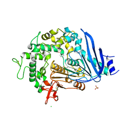
Loading
PDBj
MenuPDBj@FacebookPDBj@TwitterPDBj@YouTubewwPDB FoundationwwPDB
RCSB PDBPDBeBMRBAdv. SearchSearch help
PDB: 105 results

7AC0
DownloadVisualize
BU of 7ac0 by Molmil
Epoxide hydrolase CorEH without ligand
Descriptor: Soluble epoxide hydrolase
Authors:Palm, G.J, Lammers, M, Berndt, L.
Deposit date:2020-09-09
Release date:2021-07-21
Last modified:2024-01-31
Method:X-RAY DIFFRACTION (2.177 Å)
Cite:Promiscuous Dehalogenase Activity of the Epoxide Hydrolase CorEH from Corynebacterium sp. C12
Acs Catalysis, 11, 2021
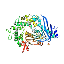
7ATL
DownloadVisualize
BU of 7atl by Molmil
EstCE1, a hydrolase with promiscuous acyltransferase activity
Descriptor: 1,2-ETHANEDIOL, DI(HYDROXYETHYL)ETHER, Esterase, ...
Authors:Palm, G.J, Lammers, M, Berndt, L.
Deposit date:2020-10-30
Release date:2020-11-18
Last modified:2024-01-31
Method:X-RAY DIFFRACTION (2.478 Å)
Cite:Discovery and Design of Family VIII Carboxylesterases as Highly Efficient Acyltransferases.
Angew.Chem.Int.Ed.Engl., 60, 2021
6Y9K
DownloadVisualize
BU of 6y9k by Molmil
Esterase EST8 with transacylase activity
Descriptor: Esterase Est8
Authors:Palm, G.J, Lammers, M, Berndt, L.
Deposit date:2020-03-09
Release date:2020-07-29
Last modified:2024-01-24
Method:X-RAY DIFFRACTION (2.298 Å)
Cite:Sequence-Based Prediction of Promiscuous Acyltransferase Activity in Hydrolases.
Angew.Chem.Int.Ed.Engl., 59, 2020
7P97
DownloadVisualize
BU of 7p97 by Molmil
Structure of 3-phospho-D-glycerate guanylyltransferase with product 3-GPPG bound
Descriptor: 3-(guanosine-5'-diphospho)-D-glycerate, 3-phospho-D-glycerate guanylyltransferase, CHLORIDE ION, ...
Authors:Palm, G.J, Berndt, L, Lammers, M.
Deposit date:2021-07-26
Release date:2022-02-02
Last modified:2024-01-31
Method:X-RAY DIFFRACTION (2.35 Å)
Cite:Diversification by CofC and Control by CofD Govern Biosynthesis and Evolution of Coenzyme F 420 and Its Derivative 3PG-F 420.
Mbio, 13, 2022
8B7Z
DownloadVisualize
BU of 8b7z by Molmil
Bacterial chalcone isomerase H33A with taxifolin
Descriptor: (2R,3R)-2-(3,4-DIHYDROXYPHENYL)-3,5,7-TRIHYDROXY-2,3-DIHYDRO-4H-CHROMEN-4-ONE, 4-(2-HYDROXYETHYL)-1-PIPERAZINE ETHANESULFONIC ACID, CHLORIDE ION, ...
Authors:Palm, G.J, Hinrichs, W.
Deposit date:2022-10-03
Release date:2022-11-23
Last modified:2024-01-31
Method:X-RAY DIFFRACTION (3 Å)
Cite:Structural Basis for (2 R ,3 R )-Taxifolin Binding and Reaction Products to the Bacterial Chalcone Isomerase of Eubacterium ramulus .
Molecules, 27, 2022
1EMC
DownloadVisualize
BU of 1emc by Molmil
GREEN FLUORESCENT PROTEIN FROM AEQUOREA VICTORIA, MUTANT
Descriptor: GREEN FLUORESCENT PROTEIN
Authors:Palm, G, Zdanov, A, Wlodawer, A.
Deposit date:1997-03-31
Release date:1997-08-20
Last modified:2023-11-15
Method:X-RAY DIFFRACTION (2.3 Å)
Cite:The structural basis for spectral variations in green fluorescent protein.
Nat.Struct.Biol., 4, 1997
2EMN
DownloadVisualize
BU of 2emn by Molmil
GREEN FLUORESCENT PROTEIN FROM AEQUOREA VICTORIA, MUTANT
Descriptor: GREEN FLUORESCENT PROTEIN
Authors:Palm, G, Zdanov, A, Wlodawer, A.
Deposit date:1997-03-31
Release date:1997-08-20
Last modified:2023-08-09
Method:X-RAY DIFFRACTION (2.3 Å)
Cite:The structural basis for spectral variations in green fluorescent protein.
Nat.Struct.Biol., 4, 1997
2EMO
DownloadVisualize
BU of 2emo by Molmil
GREEN FLUORESCENT PROTEIN FROM AEQUOREA VICTORIA, MUTANT
Descriptor: GREEN FLUORESCENT PROTEIN
Authors:Palm, G, Zdanov, A, Wlodawer, A.
Deposit date:1997-03-31
Release date:1997-08-20
Last modified:2023-08-09
Method:X-RAY DIFFRACTION (2.6 Å)
Cite:The structural basis for spectral variations in green fluorescent protein.
Nat.Struct.Biol., 4, 1997
2EMD
DownloadVisualize
BU of 2emd by Molmil
GREEN FLUORESCENT PROTEIN FROM AEQUOREA VICTORIA, MUTANT
Descriptor: GREEN FLUORESCENT PROTEIN
Authors:Palm, G, Zdanov, A, Wlodawer, A.
Deposit date:1997-03-31
Release date:1997-08-20
Last modified:2023-08-09
Method:X-RAY DIFFRACTION (2 Å)
Cite:The structural basis for spectral variations in green fluorescent protein.
Nat.Struct.Biol., 4, 1997
1EML
DownloadVisualize
BU of 1eml by Molmil
GREEN FLUORESCENT PROTEIN FROM AEQUOREA VICTORIA, MUTANT
Descriptor: GREEN FLUORESCENT PROTEIN
Authors:Palm, G, Zdanov, A, Wlodawer, A.
Deposit date:1997-03-31
Release date:1997-08-20
Last modified:2023-11-15
Method:X-RAY DIFFRACTION (2.3 Å)
Cite:The structural basis for spectral variations in green fluorescent protein.
Nat.Struct.Biol., 4, 1997
1EMK
DownloadVisualize
BU of 1emk by Molmil
GREEN FLUORESCENT PROTEIN FROM AEQUOREA VICTORIA, MUTANT
Descriptor: GREEN FLUORESCENT PROTEIN
Authors:Palm, G, Zdanov, A, Wlodawer, A.
Deposit date:1997-03-31
Release date:1997-08-20
Last modified:2023-08-09
Method:X-RAY DIFFRACTION (2.1 Å)
Cite:The structural basis for spectral variations in green fluorescent protein.
Nat.Struct.Biol., 4, 1997
1EMM
DownloadVisualize
BU of 1emm by Molmil
GREEN FLUORESCENT PROTEIN FROM AEQUOREA VICTORIA, MUTANT
Descriptor: GREEN FLUORESCENT PROTEIN
Authors:Palm, G, Zdanov, A, Wlodawer, A.
Deposit date:1997-03-31
Release date:1997-08-20
Last modified:2023-11-15
Method:X-RAY DIFFRACTION (2.3 Å)
Cite:The structural basis for spectral variations in green fluorescent protein.
Nat.Struct.Biol., 4, 1997
1EME
DownloadVisualize
BU of 1eme by Molmil
GREEN FLUORESCENT PROTEIN FROM AEQUOREA VICTORIA, MUTANT
Descriptor: GREEN FLUORESCENT PROTEIN
Authors:Palm, G, Zdanov, A, Wlodawer, A.
Deposit date:1997-03-31
Release date:1997-08-20
Last modified:2023-11-15
Method:X-RAY DIFFRACTION (2.5 Å)
Cite:The structural basis for spectral variations in green fluorescent protein.
Nat.Struct.Biol., 4, 1997
1EMF
DownloadVisualize
BU of 1emf by Molmil
GREEN FLUORESCENT PROTEIN FROM AEQUOREA VICTORIA, MUTANT
Descriptor: GREEN FLUORESCENT PROTEIN
Authors:Palm, G, Zdanov, A, Wlodawer, A.
Deposit date:1997-03-31
Release date:1997-08-20
Last modified:2023-08-09
Method:X-RAY DIFFRACTION (2.4 Å)
Cite:The structural basis for spectral variations in green fluorescent protein.
Nat.Struct.Biol., 4, 1997
6QG9
DownloadVisualize
BU of 6qg9 by Molmil
Crystal structure of Ideonella sakaiensis MHETase
Descriptor: 1,2-ETHANEDIOL, ACETATE ION, CALCIUM ION, ...
Authors:Palm, G.J, Reisky, L, Boettcher, D, Mueller, H, Michels, E.A.P, Walczak, C, Berndt, L, Weiss, M.S, Bornscheuer, U.T, Weber, G.
Deposit date:2019-01-10
Release date:2019-04-03
Last modified:2024-01-24
Method:X-RAY DIFFRACTION (2.05 Å)
Cite:Structure of the plastic-degrading Ideonella sakaiensis MHETase bound to a substrate.
Nat Commun, 10, 2019
6QGB
DownloadVisualize
BU of 6qgb by Molmil
Crystal structure of Ideonella sakaiensis MHETase bound to benzoic acid
Descriptor: (4S)-2-METHYL-2,4-PENTANEDIOL, BENZOIC ACID, CALCIUM ION, ...
Authors:Palm, G.J, Reisky, L, Boettcher, D, Mueller, H, Michels, E.A.P, Walczak, C, Berndt, L, Weiss, M.S, Bornscheuer, U.T, Weber, G.
Deposit date:2019-01-10
Release date:2019-04-03
Last modified:2019-04-24
Method:X-RAY DIFFRACTION (2.2 Å)
Cite:Structure of the plastic-degrading Ideonella sakaiensis MHETase bound to a substrate.
Nat Commun, 10, 2019
6QGA
DownloadVisualize
BU of 6qga by Molmil
Crystal structure of Ideonella sakaiensis MHETase bound to the non-hydrolyzable ligand MHETA
Descriptor: (4S)-2-METHYL-2,4-PENTANEDIOL, 4-(2-hydroxyethylcarbamoyl)benzoic acid, CALCIUM ION, ...
Authors:Palm, G.J, Reisky, L, Boettcher, D, Mueller, H, Michels, E.A.P, Walczak, C, Berndt, L, Weiss, M.S, Bornscheuer, U.T, Weber, G.
Deposit date:2019-01-10
Release date:2019-04-03
Last modified:2024-01-24
Method:X-RAY DIFFRACTION (2.1 Å)
Cite:Structure of the plastic-degrading Ideonella sakaiensis MHETase bound to a substrate.
Nat Commun, 10, 2019
6QGC
DownloadVisualize
BU of 6qgc by Molmil
PETase from Ideonella sakaiensis without ligand
Descriptor: CHLORIDE ION, Poly(ethylene terephthalate) hydrolase, SULFATE ION
Authors:Palm, G.J, Reisky, L, Boettcher, D, Mueller, H, Michels, E.A.P, Walczak, C, Berndt, L, Weiss, M.S, Bornscheuer, U.T, Weber, G.
Deposit date:2019-01-10
Release date:2019-04-03
Last modified:2024-01-24
Method:X-RAY DIFFRACTION (2 Å)
Cite:Structure of the plastic-degrading Ideonella sakaiensis MHETase bound to a substrate.
Nat Commun, 10, 2019
6GYI
DownloadVisualize
BU of 6gyi by Molmil
Azurin fom Pseudomonas aeruginosa treated with hydrosulfide
Descriptor: Azurin, CALCIUM ION, COPPER (II) ION
Authors:Palm, G.J, Kohlhause, D.
Deposit date:2018-06-29
Release date:2019-01-30
Last modified:2024-01-17
Method:X-RAY DIFFRACTION (1.6 Å)
Cite:Azurin and HS-: towards implementation of a sensor for HS-detection.
Eur J Inorg Chem, 2019
6IAV
DownloadVisualize
BU of 6iav by Molmil
CO-AZURIN FROM PSEUDOMONAS AERUGINOSA TREATED WITH HYDROSULFIDE
Descriptor: Azurin, CALCIUM ION, COBALT (II) ION
Authors:Palm, G.J, Kohlhause, D.
Deposit date:2018-11-27
Release date:2019-01-30
Last modified:2024-01-24
Method:X-RAY DIFFRACTION (2 Å)
Cite:Azurin and HS-: towards implementation of a sensor for HS- detection.
Eur J Inorg Chem, 2019
4CUO
DownloadVisualize
BU of 4cuo by Molmil
Banyan peroxidase with glycosylation
Descriptor: 2-acetamido-2-deoxy-beta-D-glucopyranose, BANYAN PEROXIDASE, CALCIUM ION, ...
Authors:Palm, G.J, Sharma, A, Hinrichs, W.
Deposit date:2014-03-20
Release date:2014-07-23
Last modified:2023-11-15
Method:X-RAY DIFFRACTION (1.67 Å)
Cite:Post-Translational Modification and Extended Glycosylation Pattern of a Plant Latex Peroxidase of Native Source Characterized by X-Ray Crystallography.
FEBS J., 281, 2014
2WUR
DownloadVisualize
BU of 2wur by Molmil
Atomic resolution structure of GFP measured on a rotating anode
Descriptor: ETHANOL, GREEN FLUORESCENT PROTEIN, ISOPROPYL ALCOHOL
Authors:Palm, G.J, Schierbeek, A.J, Kloos, M.
Deposit date:2009-10-07
Release date:2010-05-12
Last modified:2023-11-15
Method:X-RAY DIFFRACTION (0.9 Å)
Cite:Visualizing Proton Antenna in a High-Resolution Green Fluorescent Protein Structure.
J.Am.Chem.Soc., 132, 2010
1QMH
DownloadVisualize
BU of 1qmh by Molmil
Crystal structure of RNA 3'-terminal phosphate cyclase, an ubiquitous enzyme with unusual topology
Descriptor: 1-HYDROXYSULFANYL-4-MERCAPTO-BUTANE-2,3-DIOL, CITRIC ACID, RNA 3'-TERMINAL PHOSPHATE CYCLASE
Authors:Palm, G.J, Billy, E, Filipowicz, W, Wlodawer, A.
Deposit date:1999-09-28
Release date:2000-01-11
Last modified:2023-12-13
Method:X-RAY DIFFRACTION (2.1 Å)
Cite:Crystal Structure of RNA 3'-Terminal Phosphate Cyclase, a Ubiquitous Enzyme with Unusual Topology
Structure, 8, 2000
4AL2
DownloadVisualize
BU of 4al2 by Molmil
peptide deformylase (Ni-form) with hydrosulfide
Descriptor: HYDROSULFURIC ACID, NICKEL (II) ION, PEPTIDE DEFORMYLASE
Authors:Palm, G.J, Hinrichs, W.
Deposit date:2012-02-29
Release date:2012-04-04
Last modified:2023-12-20
Method:X-RAY DIFFRACTION (2.6 Å)
Cite:A Fret Enzyme-Based Probe for Monitoring Hydrogen Sulfide.
Inorg.Chem., 51, 2012
4ECA
DownloadVisualize
BU of 4eca by Molmil
ASPARAGINASE FROM E. COLI, MUTANT T89V WITH COVALENTLY BOUND ASPARTATE
Descriptor: L-ASPARAGINE AMIDOHYDROLASE
Authors:Palm, G.J, Lubkowski, J, Wlodawer, A.
Deposit date:1997-02-21
Release date:1997-06-16
Last modified:2023-08-09
Method:X-RAY DIFFRACTION (2.2 Å)
Cite:A covalently bound catalytic intermediate in Escherichia coli asparaginase: crystal structure of a Thr-89-Val mutant.
FEBS Lett., 390, 1996

220472

PDB entries from 2024-05-29

PDB statisticsPDBj update infoContact PDBjnumon