Loading
PDBj
MenuPDBj@FacebookPDBj@TwitterPDBj@YouTubewwPDB FoundationwwPDB
RCSB PDBPDBeBMRBAdv. SearchSearch help
PDB: 7 results

6I41
DownloadVisualize
BU of 6i41 by Molmil
Co-crystal structure of human SPOP MATH domain (wild-type) and human BRD3 fragment
Descriptor: Bromodomain-containing protein 3, Speckle-type POZ protein
Authors:Ostertag, M.S, Popowicz, G.M, Sattler, M.
Deposit date:2018-11-08
Release date:2019-05-08
Last modified:2024-01-24
Method:X-RAY DIFFRACTION (1.9 Å)
Cite:Structural Insights into BET Client Recognition of Endometrial and Prostate Cancer-Associated SPOP Mutants.
J.Mol.Biol., 431, 2019
6I5P
DownloadVisualize
BU of 6i5p by Molmil
Co-crystal structure of human SPOP MATH domain (E47K) and human BRD3 fragment
Descriptor: Bromodomain-containing protein 3, Speckle-type POZ protein
Authors:Ostertag, M.S, Popowicz, G.M, Sattler, M.
Deposit date:2018-11-14
Release date:2019-05-08
Last modified:2024-01-24
Method:X-RAY DIFFRACTION (1.81 Å)
Cite:Structural Insights into BET Client Recognition of Endometrial and Prostate Cancer-Associated SPOP Mutants.
J.Mol.Biol., 431, 2019
6I68
DownloadVisualize
BU of 6i68 by Molmil
Co-crystal structure of human SPOP MATH domain (M117V) and human BRD3 fragment
Descriptor: Bromodomain-containing protein 3, Speckle-type POZ protein
Authors:Ostertag, M.S, Popowicz, G.M, Sattler, M.
Deposit date:2018-11-15
Release date:2019-05-08
Last modified:2024-01-24
Method:X-RAY DIFFRACTION (1.85 Å)
Cite:Structural Insights into BET Client Recognition of Endometrial and Prostate Cancer-Associated SPOP Mutants.
J.Mol.Biol., 431, 2019
6I7A
DownloadVisualize
BU of 6i7a by Molmil
Co-crystal structure of human SPOP MATH domain (D140N) and human BRD3 fragment
Descriptor: Bromodomain-containing protein 3, Speckle-type POZ protein
Authors:Ostertag, M.S, Popowicz, G.M, Sattler, M.
Deposit date:2018-11-16
Release date:2019-05-08
Last modified:2024-01-24
Method:X-RAY DIFFRACTION (2.2 Å)
Cite:Structural Insights into BET Client Recognition of Endometrial and Prostate Cancer-Associated SPOP Mutants.
J.Mol.Biol., 431, 2019
6F8G
DownloadVisualize
BU of 6f8g by Molmil
Co-crystal structure of SPOP MATH domain and hamster Pdx1 fragment
Descriptor: Pancreas/duodenum homeobox protein 1, Speckle-type POZ protein
Authors:Ostertag, M.S, Popowicz, G.M, Sattler, M.
Deposit date:2017-12-13
Release date:2018-11-28
Last modified:2024-01-17
Method:X-RAY DIFFRACTION (2.03 Å)
Cite:The Structure of the SPOP-Pdx1 Interface Reveals Insights into the Phosphorylation-Dependent Binding Regulation.
Structure, 27, 2019
6F8F
DownloadVisualize
BU of 6f8f by Molmil
Co-crystal structure of SPOP MATH domain and human Pdx1 fragment
Descriptor: Pancreas/duodenum homeobox protein 1, Speckle-type POZ protein
Authors:Ostertag, M.S, Popowicz, G.M, Sattler, M.
Deposit date:2017-12-13
Release date:2018-11-28
Last modified:2024-01-17
Method:X-RAY DIFFRACTION (2 Å)
Cite:The Structure of the SPOP-Pdx1 Interface Reveals Insights into the Phosphorylation-Dependent Binding Regulation.
Structure, 27, 2019
6SPT
DownloadVisualize
BU of 6spt by Molmil
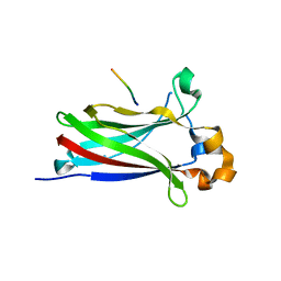
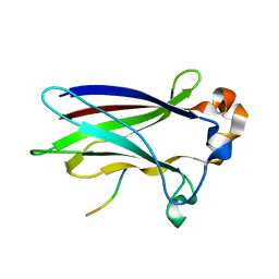
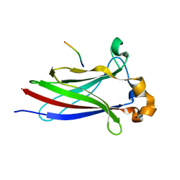
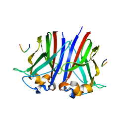
High resolution crystal structure of N-terminal domain of PEX14 from Trypanosoma brucei in complex with the fist compound with sub-micromolar trypanocidal activity
Descriptor: 5-[(4-methoxynaphthalen-1-yl)methyl]-1-[2-[(2-methyl-1-oxidanyl-propan-2-yl)amino]ethyl]-~{N}-(naphthalen-1-ylmethyl)-6,7-dihydro-4~{H}-pyrazolo[4,3-c]pyridine-3-carboxamide, BETA-MERCAPTOETHANOL, CHLORIDE ION, ...
Authors:Napolitano, V, Dawidowski, M, Kalel, V.C, Fino, R, Emmanouilidis, L, Lenhart, D, Ostertag, M, Kaiser, M, Kolonko, M, Schilebs, W, Maser, P, Tetko, I, Hadian, K, Plettenburg, O, Erdmann, R, Sattler, M, Popowicz, G.M, Dubin, G.
Deposit date:2019-09-02
Release date:2020-01-01
Last modified:2020-02-05
Method:X-RAY DIFFRACTION (1.2 Å)
Cite:Structure-Activity Relationship in Pyrazolo[4,3-c]pyridines, First Inhibitors of PEX14-PEX5 Protein-Protein Interaction with Trypanocidal Activity.
J.Med.Chem., 63, 2020

219140

PDB entries from 2024-05-01

PDB statisticsPDBj update infoContact PDBjnumon