Loading
PDBj
MenuPDBj@FacebookPDBj@TwitterPDBj@YouTubewwPDB FoundationwwPDB
RCSB PDBPDBeBMRBAdv. SearchSearch help
PDB: 122 results

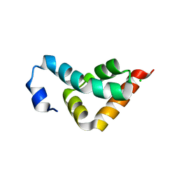
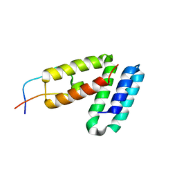
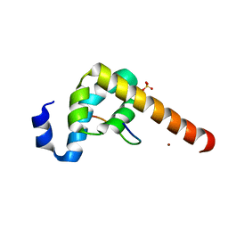
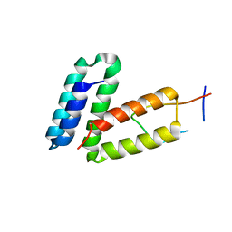
4G3O
DownloadVisualize
BU of 4g3o by Molmil
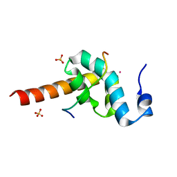
Crystal structure of the CUE domain of the E3 ubiquitin ligase AMFR (gp78)
Descriptor: E3 ubiquitin-protein ligase AMFR
Authors:Kozlov, G, LePage, K, Gehring, K.
Deposit date:2012-07-15
Release date:2012-07-25
Last modified:2023-09-13
Method:X-RAY DIFFRACTION (1.6 Å)
Cite:Crystal structure of the CUE domain of the E3 ubiquitin ligase AMFR (gp78)
To be Published
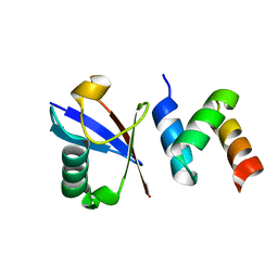
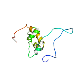
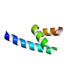
4I6X
DownloadVisualize
BU of 4i6x by Molmil
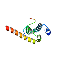
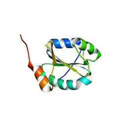
Crystal Structure of Non-catalyic Domain of Protein Disulfide Isomerase-related (PDIr) Protein
Descriptor: Protein disulfide-isomerase A5
Authors:Kozlov, G, Vinaik, R, Gehring, K.
Deposit date:2012-11-30
Release date:2013-04-03
Last modified:2013-05-08
Method:X-RAY DIFFRACTION (1.5 Å)
Cite:Structure of the Non-Catalytic Domain of the Protein Disulfide Isomerase-Related Protein (PDIR) Reveals Function in Protein Binding.
Plos One, 8, 2013
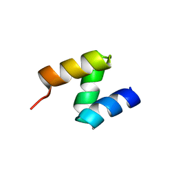
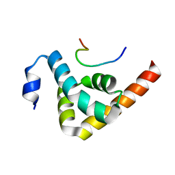
3KUR
DownloadVisualize
BU of 3kur by Molmil
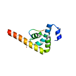
Crystal structure of the MLLE domain of poly(A)-binding protein
Descriptor: CHLORIDE ION, Polyadenylate-binding protein 1
Authors:Kozlov, G, Gehring, K.
Deposit date:2009-11-27
Release date:2010-02-09
Last modified:2023-09-06
Method:X-RAY DIFFRACTION (2.5 Å)
Cite:Molecular Determinants of PAM2 Recognition by the MLLE Domain of Poly(A)-Binding Protein.
J.Mol.Biol., 397, 2010
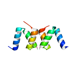
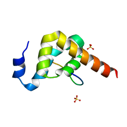
2OOB
DownloadVisualize
BU of 2oob by Molmil
crystal structure of the UBA domain from Cbl-b ubiquitin ligase in complex with ubiquitin
Descriptor: E3 ubiquitin-protein ligase CBL-B, Ubiquitin
Authors:Kozlov, G, Gehring, K.
Deposit date:2007-01-25
Release date:2007-02-06
Last modified:2023-08-30
Method:X-RAY DIFFRACTION (1.9 Å)
Cite:Structural basis for ubiquitin-mediated dimerization and activation of the ubiquitin protein ligase Cbl-b.
Mol.Cell, 27, 2007
2OOA
DownloadVisualize
BU of 2ooa by Molmil
crystal structure of the UBA domain from Cbl-b ubiquitin ligase
Descriptor: E3 ubiquitin-protein ligase CBL-B
Authors:Kozlov, G, Gehring, K.
Deposit date:2007-01-25
Release date:2007-02-06
Last modified:2023-12-27
Method:X-RAY DIFFRACTION (1.56 Å)
Cite:Structural basis for ubiquitin-mediated dimerization and activation of the ubiquitin protein ligase Cbl-b.
Mol.Cell, 27, 2007
2OO9
DownloadVisualize
BU of 2oo9 by Molmil
crystal structure of the UBA domain from human c-Cbl ubiquitin ligase
Descriptor: E3 ubiquitin-protein ligase CBL
Authors:Kozlov, G, Gehring, K.
Deposit date:2007-01-25
Release date:2007-02-06
Last modified:2023-12-27
Method:X-RAY DIFFRACTION (2.1 Å)
Cite:Structural basis for UBA-mediated dimerization of c-Cbl ubiquitin ligase.
J.Biol.Chem., 282, 2007
3KTR
DownloadVisualize
BU of 3ktr by Molmil
Structural basis of ataxin-2 recognition by poly(A)-binding protein
Descriptor: Ataxin-2, CADMIUM ION, Polyadenylate-binding protein 1, ...
Authors:Kozlov, G, Gehring, K.
Deposit date:2009-11-25
Release date:2010-02-23
Last modified:2023-09-06
Method:X-RAY DIFFRACTION (1.7 Å)
Cite:Structural basis of binding of P-body-associated proteins GW182 and ataxin-2 by the Mlle domain of poly(A)-binding protein.
J.Biol.Chem., 285, 2010
3KUT
DownloadVisualize
BU of 3kut by Molmil
Crystal structure of the MLLE domain of poly(A)-binding protein in complex with the binding region of Paip2
Descriptor: CHLORIDE ION, PAIP2 protein, Polyadenylate-binding protein 1
Authors:Kozlov, G, Gehring, K.
Deposit date:2009-11-27
Release date:2010-02-09
Last modified:2023-09-06
Method:X-RAY DIFFRACTION (1.5 Å)
Cite:Molecular Determinants of PAM2 Recognition by the MLLE Domain of Poly(A)-Binding Protein.
J.Mol.Biol., 397, 2010
3KUS
DownloadVisualize
BU of 3kus by Molmil
Crystal structure of the MLLE domain of poly(A)-binding protein in complex with the binding region of Paip2
Descriptor: 4-(2-HYDROXYETHYL)-1-PIPERAZINE ETHANESULFONIC ACID, GLYCEROL, PAIP2 protein, ...
Authors:Kozlov, G, Gehring, K.
Deposit date:2009-11-27
Release date:2010-02-09
Last modified:2023-09-06
Method:X-RAY DIFFRACTION (1.4 Å)
Cite:Molecular Determinants of PAM2 Recognition by the MLLE Domain of Poly(A)-Binding Protein.
J.Mol.Biol., 397, 2010
1GH8
DownloadVisualize
BU of 1gh8 by Molmil
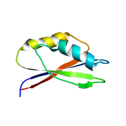
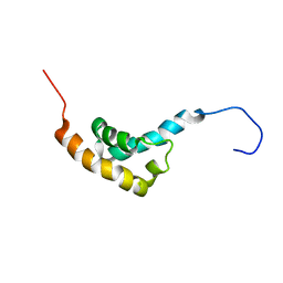
SOLUTION STRUCTURE OF THE ARCHAEAL TRANSLATION ELONGATION FACTOR 1BETA FROM METHANOBACTERIUM THERMOAUTOTROPHICUM
Descriptor: TRANSLATION ELONGATION FACTOR 1BETA
Authors:Kozlov, G, Ekiel, I, Gehring, K, Northeast Structural Genomics Consortium (NESG)
Deposit date:2000-11-30
Release date:2000-12-13
Last modified:2023-12-27
Method:SOLUTION NMR
Cite:Rapid fold and structure determination of the archaeal translation elongation factor 1beta from Methanobacterium thermoautotrophicum.
J.Biomol.NMR, 17, 2000
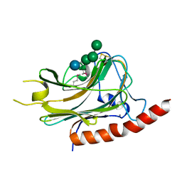
3O0X
DownloadVisualize
BU of 3o0x by Molmil
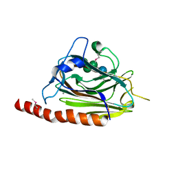
Structural basis of carbohydrate recognition by calreticulin
Descriptor: CALCIUM ION, Calreticulin, alpha-D-glucopyranose-(1-3)-alpha-D-mannopyranose-(1-2)-alpha-D-mannopyranose-(1-2)-alpha-D-mannopyranose
Authors:Kozlov, G, Gehring, K.
Deposit date:2010-07-20
Release date:2010-09-29
Last modified:2023-12-06
Method:X-RAY DIFFRACTION (2.01 Å)
Cite:Structural basis of carbohydrate recognition by calreticulin.
J.Biol.Chem., 285, 2010
3O10
DownloadVisualize
BU of 3o10 by Molmil
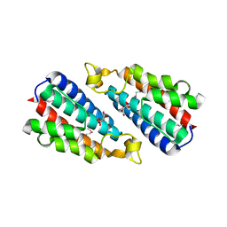
Crystal structure of the HEPN domain from human sacsin
Descriptor: MALONATE ION, Sacsin
Authors:Kozlov, G, Gehring, K.
Deposit date:2010-07-20
Release date:2011-03-30
Last modified:2014-07-30
Method:X-RAY DIFFRACTION (1.9 Å)
Cite:Structural basis of defects in the sacsin HEPN domain responsible for autosomal recessive spastic ataxia of Charlevoix-Saguenay (ARSACS).
J.Biol.Chem., 286, 2011
5TDC
DownloadVisualize
BU of 5tdc by Molmil
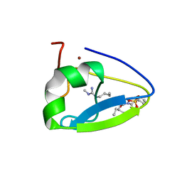
Crystal structure of the human UBR-box domain from UBR1 in complex with monomethylated arginine peptide.
Descriptor: E3 ubiquitin-protein ligase UBR1, NMM-ILE-PHE-SER peptide, SULFATE ION, ...
Authors:Kozlov, G, Munoz-Escobar, J, Matta-Camacho, E, Gehring, K.
Deposit date:2016-09-19
Release date:2017-03-22
Last modified:2023-10-04
Method:X-RAY DIFFRACTION (1.607 Å)
Cite:Bound Waters Mediate Binding of Diverse Substrates to a Ubiquitin Ligase.
Structure, 25, 2017
5TSR
DownloadVisualize
BU of 5tsr by Molmil
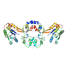
Crystal structure of PRL-3 phosphatase in complex with the Bateman domain of CNNM3 magnesium transporter
Descriptor: Metal transporter CNNM3, Protein tyrosine phosphatase type IVA 3
Authors:Kozlov, G, Zhang, H, Gehring, K.
Deposit date:2016-10-31
Release date:2017-02-08
Last modified:2023-10-04
Method:X-RAY DIFFRACTION (3.188 Å)
Cite:PRL3 phosphatase active site is required for binding the putative magnesium transporter CNNM3.
Sci Rep, 7, 2017
5V8Z
DownloadVisualize
BU of 5v8z by Molmil
Crystal structure of ERp29 D-domain in complex with the P-domain of calmegin
Descriptor: Calmegin, Endoplasmic reticulum resident protein 29
Authors:Kozlov, G, Munoz-Escobar, J, Gehring, K.
Deposit date:2017-03-22
Release date:2017-06-21
Last modified:2023-10-04
Method:X-RAY DIFFRACTION (2.105 Å)
Cite:Mapping the ER Interactome: The P Domains of Calnexin and Calreticulin as Plurivalent Adapters for Foldases and Chaperones.
Structure, 25, 2017
1G9L
DownloadVisualize
BU of 1g9l by Molmil
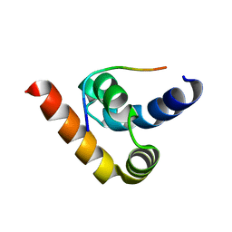
SOLUTION STRUCTURE OF THE PABC DOMAIN OF HUMAN POLY(A) BINDING PROTEIN
Descriptor: POLYADENYLATE-BINDING PROTEIN 1
Authors:Kozlov, G, Trempe, J.-F, Khaleghpour, K, Kahvejian, A, Ekiel, I, Gehring, K.
Deposit date:2000-11-24
Release date:2001-03-14
Last modified:2024-05-22
Method:SOLUTION NMR
Cite:Structure and function of the C-terminal PABC domain of human poly(A)-binding protein.
Proc.Natl.Acad.Sci.USA, 98, 2001
3KTP
DownloadVisualize
BU of 3ktp by Molmil
Structural basis of GW182 recognition by poly(A)-binding protein
Descriptor: Polyadenylate-binding protein 1, Trinucleotide repeat-containing gene 6C protein
Authors:Kozlov, G, Gehring, K.
Deposit date:2009-11-25
Release date:2010-02-23
Last modified:2023-09-06
Method:X-RAY DIFFRACTION (1.5 Å)
Cite:Structural basis of binding of P-body-associated proteins GW182 and ataxin-2 by the Mlle domain of poly(A)-binding protein.
J.Biol.Chem., 285, 2010
3KUJ
DownloadVisualize
BU of 3kuj by Molmil
Crystal structure of C-terminal domain of PABPC1 in complex with binding region of eRF3a
Descriptor: GSPT1 protein, Polyadenylate-binding protein 1, SULFATE ION
Authors:Kozlov, G, Gehring, K.
Deposit date:2009-11-27
Release date:2010-05-12
Last modified:2023-09-06
Method:X-RAY DIFFRACTION (1.4 Å)
Cite:Molecular basis of eRF3 recognition by the MLLE domain of poly(A)-binding protein.
Plos One, 5, 2010
3KUI
DownloadVisualize
BU of 3kui by Molmil
Crystal structure of C-terminal domain of PABPC1 in complex with binding region of eRF3a
Descriptor: GSPT1 protein, Polyadenylate-binding protein 1, SULFATE ION, ...
Authors:Kozlov, G, Gehring, K.
Deposit date:2009-11-27
Release date:2010-05-12
Last modified:2023-09-06
Method:X-RAY DIFFRACTION (2.3 Å)
Cite:Molecular basis of eRF3 recognition by the MLLE domain of poly(A)-binding protein.
Plos One, 5, 2010
1GH9
DownloadVisualize
BU of 1gh9 by Molmil
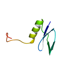
SOLUTION STRUCTURE OF A 8.3 KDA PROTEIN (GENE MTH1184) FROM METHANOBACTERIUM THERMOAUTOTROPHICUM
Descriptor: 8.3 KDA PROTEIN (GENE MTH1184)
Authors:Kozlov, G, Ekiel, I, Gehring, K, Northeast Structural Genomics Consortium (NESG)
Deposit date:2000-11-30
Release date:2000-12-11
Last modified:2023-12-27
Method:SOLUTION NMR
Cite:Structural proteomics of an archaeon.
Nat.Struct.Biol., 7, 2000
1IFW
DownloadVisualize
BU of 1ifw by Molmil
SOLUTION STRUCTURE OF C-TERMINAL DOMAIN OF POLY(A) BINDING PROTEIN FROM SACCHAROMYCES CEREVISIAE
Descriptor: POLYADENYLATE-BINDING PROTEIN, CYTOPLASMIC AND NUCLEAR
Authors:Kozlov, G, Siddiqui, N, Coillet-Matillon, S, Sprules, T, Ekiel, I, Gehring, K.
Deposit date:2001-04-13
Release date:2002-07-24
Last modified:2024-05-22
Method:SOLUTION NMR
Cite:Solution structure of the orphan PABC domain from Saccharomyces cerevisiae poly(A)-binding protein.
J.Biol.Chem., 277, 2002
3NTW
DownloadVisualize
BU of 3ntw by Molmil
Structure of the MLLE domain of EDD in complex with a PAM2 peptide from Paip1
Descriptor: E3 ubiquitin-protein ligase UBR5, Polyadenylate-binding protein-interacting protein 1
Authors:Kozlov, G, Gehring, K.
Deposit date:2010-07-05
Release date:2011-07-06
Last modified:2023-09-06
Method:X-RAY DIFFRACTION (2.6 Å)
Cite:The MLLE domain of the ubiquitin ligase UBR5 binds to its catalytic domain to regulate substrate binding.
J. Biol. Chem., 290, 2015
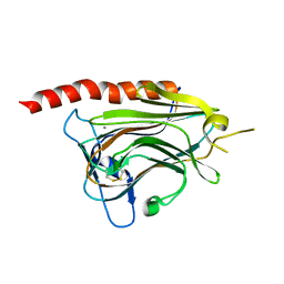
3O0W
DownloadVisualize
BU of 3o0w by Molmil
Structural basis of carbohydrate recognition by calreticulin
Descriptor: CALCIUM ION, Calreticulin, alpha-D-glucopyranose-(1-3)-alpha-D-mannopyranose-(1-2)-alpha-D-mannopyranose-(1-2)-alpha-D-mannopyranose
Authors:Kozlov, G, Gehring, K.
Deposit date:2010-07-20
Release date:2010-09-29
Last modified:2020-07-29
Method:X-RAY DIFFRACTION (1.95 Å)
Cite:Structural basis of carbohydrate recognition by calreticulin.
J.Biol.Chem., 285, 2010
3O0V
DownloadVisualize
BU of 3o0v by Molmil
Crystal structure of the calreticulin lectin domain
Descriptor: CALCIUM ION, Calreticulin
Authors:Kozlov, G, Gehring, K.
Deposit date:2010-07-20
Release date:2010-09-29
Last modified:2023-09-06
Method:X-RAY DIFFRACTION (2.3 Å)
Cite:Structural basis of carbohydrate recognition by calreticulin.
J.Biol.Chem., 285, 2010
5V90
DownloadVisualize
BU of 5v90 by Molmil
Crystal structure of ERp29 D-domain in complex with the P-domain of calreticulin
Descriptor: Calreticulin, Endoplasmic reticulum resident protein 29, GLYCEROL
Authors:Kozlov, G, Munoz-Escobar, J, Gehring, K.
Deposit date:2017-03-22
Release date:2017-06-21
Last modified:2023-10-04
Method:X-RAY DIFFRACTION (3.255 Å)
Cite:Mapping the ER Interactome: The P Domains of Calnexin and Calreticulin as Plurivalent Adapters for Foldases and Chaperones.
Structure, 25, 2017

220113

PDB entries from 2024-05-22

PDB statisticsPDBj update infoContact PDBjnumon