Loading
PDBj
MenuPDBj@FacebookPDBj@TwitterPDBj@YouTubewwPDB FoundationwwPDB
RCSB PDBPDBeBMRBAdv. SearchSearch help
PDB: 2350 results

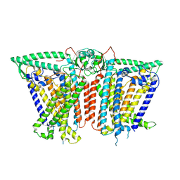
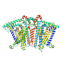
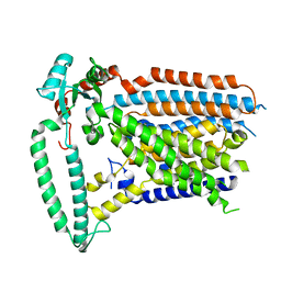
8XW1
DownloadVisualize
BU of 8xw1 by Molmil
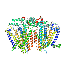
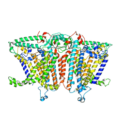
Cryo-EM structure of OSCA1.2-V335W-DDM state
Descriptor: Calcium permeable stress-gated cation channel 1
Authors:Zhang, Y, Han, Y.
Deposit date:2024-01-15
Release date:2024-04-10
Last modified:2024-05-08
Method:ELECTRON MICROSCOPY (4.49 Å)
Cite:Mechanical activation opens a lipid-lined pore in OSCA ion channels.
Nature, 628, 2024
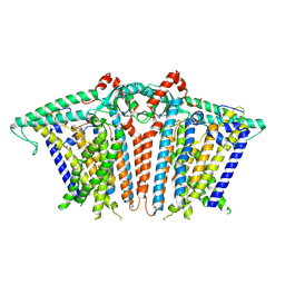
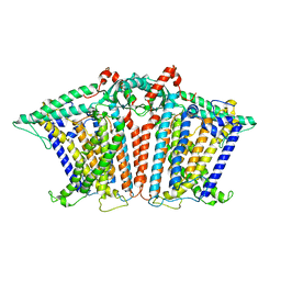
8XVY
DownloadVisualize
BU of 8xvy by Molmil
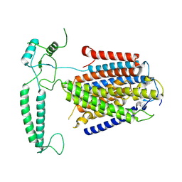
Cryo-EM structure of OSCA3.1-2E(R611E-R619E)-closed/open state
Descriptor: CSC1-like protein ERD4
Authors:Zhang, Y, Han, Y.
Deposit date:2024-01-15
Release date:2024-04-10
Last modified:2024-05-08
Method:ELECTRON MICROSCOPY (3.71 Å)
Cite:Mechanical activation opens a lipid-lined pore in OSCA ion channels.
Nature, 628, 2024
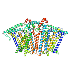
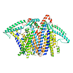
8XW3
DownloadVisualize
BU of 8xw3 by Molmil
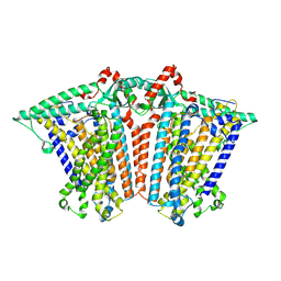
Cryo-EM structure of OSCA1.2-DOPC-1:50-expanded state
Descriptor: Calcium permeable stress-gated cation channel 1
Authors:Zhang, Y, Han, Y.
Deposit date:2024-01-15
Release date:2024-04-10
Last modified:2024-05-08
Method:ELECTRON MICROSCOPY (3.63 Å)
Cite:Mechanical activation opens a lipid-lined pore in OSCA ion channels.
Nature, 628, 2024
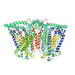
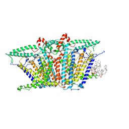
8XW2
DownloadVisualize
BU of 8xw2 by Molmil
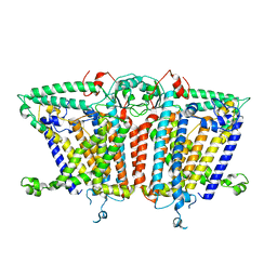
Cryo-EM structure of OSCA1.2-DOPC-1:50-contracted state
Descriptor: Calcium permeable stress-gated cation channel 1
Authors:Zhang, Y, Han, Y.
Deposit date:2024-01-15
Release date:2024-04-10
Last modified:2024-05-08
Method:ELECTRON MICROSCOPY (3.59 Å)
Cite:Mechanical activation opens a lipid-lined pore in OSCA ion channels.
Nature, 628, 2024
8XW0
DownloadVisualize
BU of 8xw0 by Molmil
Cryo-EM structure of OSCA3.1-GDN state
Descriptor: CSC1-like protein ERD4, O-[(R)-{[(2R)-2,3-bis(octadecanoyloxy)propyl]oxy}(hydroxy)phosphoryl]-L-serine
Authors:Zhang, Y, Han, Y.
Deposit date:2024-01-15
Release date:2024-04-10
Last modified:2024-05-08
Method:ELECTRON MICROSCOPY (3.11 Å)
Cite:Mechanical activation opens a lipid-lined pore in OSCA ion channels.
Nature, 628, 2024
8XVZ
DownloadVisualize
BU of 8xvz by Molmil
Cryo-EM structure of OSCA3.1-2E(R611E-R619E)-closed/'desensitized' state
Descriptor: CSC1-like protein ERD4
Authors:Zhang, Y, Han, Y.
Deposit date:2024-01-15
Release date:2024-04-10
Last modified:2024-05-08
Method:ELECTRON MICROSCOPY (3.78 Å)
Cite:Mechanical activation opens a lipid-lined pore in OSCA ion channels.
Nature, 628, 2024
8XW4
DownloadVisualize
BU of 8xw4 by Molmil
Cryo-EM structure of TMEM63B-Digitonin state
Descriptor: CSC1-like protein 2
Authors:Zhang, Y, Han, Y.
Deposit date:2024-01-15
Release date:2024-04-10
Last modified:2024-05-08
Method:ELECTRON MICROSCOPY (3.84 Å)
Cite:Mechanical activation opens a lipid-lined pore in OSCA ion channels.
Nature, 628, 2024
8XVX
DownloadVisualize
BU of 8xvx by Molmil
Cryo-EM structure of OSCA1.2-DOPC-1:20-expanded state
Descriptor: Calcium permeable stress-gated cation channel 1
Authors:Zhang, Y, Han, Y.
Deposit date:2024-01-15
Release date:2024-04-10
Last modified:2024-05-08
Method:ELECTRON MICROSCOPY (3.32 Å)
Cite:Mechanical activation opens a lipid-lined pore in OSCA ion channels.
Nature, 628, 2024
8XRY
DownloadVisualize
BU of 8xry by Molmil
Cryo-EM structure of OSCA3.1-1.1ver(Y367N-G454S-Y458I)-open/open state
Descriptor: CSC1-like protein ERD4
Authors:Zhang, Y, Han, Y.
Deposit date:2024-01-08
Release date:2024-04-10
Last modified:2024-05-08
Method:ELECTRON MICROSCOPY (3.84 Å)
Cite:Mechanical activation opens a lipid-lined pore in OSCA ion channels.
Nature, 628, 2024
8XNG
DownloadVisualize
BU of 8xng by Molmil
Cryo-EM structure of OSCA1.2-liposome-inside-out closed state
Descriptor: Calcium permeable stress-gated cation channel 1
Authors:Zhang, Y, Han, Y.
Deposit date:2023-12-29
Release date:2024-04-10
Last modified:2024-05-08
Method:ELECTRON MICROSCOPY (3.56 Å)
Cite:Mechanical activation opens a lipid-lined pore in OSCA ion channels.
Nature, 628, 2024
8XS4
DownloadVisualize
BU of 8xs4 by Molmil
Cryo-EM structure of OSCA1.2-DOPC-1:20-contracted1 state
Descriptor: Calcium permeable stress-gated cation channel 1
Authors:Zhang, Y, Han, Y.
Deposit date:2024-01-08
Release date:2024-04-10
Last modified:2024-05-08
Method:ELECTRON MICROSCOPY (3.23 Å)
Cite:Mechanical activation opens a lipid-lined pore in OSCA ion channels.
Nature, 628, 2024
8XS5
DownloadVisualize
BU of 8xs5 by Molmil
Cryo-EM structure of OSCA1.2-DOPC-1:20-contracted2 state
Descriptor: Calcium permeable stress-gated cation channel 1
Authors:Zhang, Y, Han, Y.
Deposit date:2024-01-08
Release date:2024-04-10
Last modified:2024-05-08
Method:ELECTRON MICROSCOPY (3.33 Å)
Cite:Mechanical activation opens a lipid-lined pore in OSCA ion channels.
Nature, 628, 2024
8XAJ
DownloadVisualize
BU of 8xaj by Molmil
Cryo-EM structure of OSCA1.2-liposome-inside-in open state
Descriptor: 1,2-DIOLEOYL-SN-GLYCERO-3-PHOSPHOCHOLINE, Calcium permeable stress-gated cation channel 1
Authors:Zhang, Y, Han, Y.
Deposit date:2023-12-04
Release date:2024-04-10
Last modified:2024-05-08
Method:ELECTRON MICROSCOPY (3.29 Å)
Cite:Mechanical activation opens a lipid-lined pore in OSCA ion channels.
Nature, 628, 2024
8XS0
DownloadVisualize
BU of 8xs0 by Molmil
Cryo-EM structure of OSCA3.1-1.1ver(Y367N-G454S-Y458I)-open/'desensitized' state
Descriptor: CSC1-like protein ERD4
Authors:Zhang, Y, Han, Y.
Deposit date:2024-01-08
Release date:2024-04-10
Last modified:2024-05-08
Method:ELECTRON MICROSCOPY (3.89 Å)
Cite:Mechanical activation opens a lipid-lined pore in OSCA ion channels.
Nature, 628, 2024
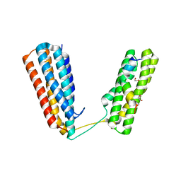
6CB7
DownloadVisualize
BU of 6cb7 by Molmil
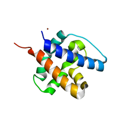
CRYSTAL STRUCTURE OF VACCINIA VIRUS A6 N-TERMINUS (SPACE GROUP C2)
Descriptor: NICKEL (II) ION, Protein A6
Authors:Han, Y, Zhang, B, Deng, J.
Deposit date:2018-02-02
Release date:2018-12-12
Last modified:2023-10-04
Method:X-RAY DIFFRACTION (1.6 Å)
Cite:Structure of a lipid-bound viral membrane assembly protein reveals a modality for enclosing the lipid bilayer.
Proc. Natl. Acad. Sci. U.S.A., 115, 2018
6CB6
DownloadVisualize
BU of 6cb6 by Molmil
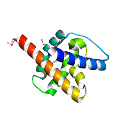
CRYSTAL STRUCTURE OF VACCINIA VIRUS A6 N-TERMINUS (SPACE GROUP C2)
Descriptor: Protein A6
Authors:Han, Y, Zhang, B, Deng, J.
Deposit date:2018-02-02
Release date:2018-12-12
Last modified:2024-10-23
Method:X-RAY DIFFRACTION (1.8 Å)
Cite:Structure of a lipid-bound viral membrane assembly protein reveals a modality for enclosing the lipid bilayer.
Proc. Natl. Acad. Sci. U.S.A., 115, 2018
3QH2
DownloadVisualize
BU of 3qh2 by Molmil
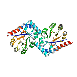
Crystal structure of TenI from Bacillus subtilis complexed with product cThz-P
Descriptor: 4-methyl-5-[2-(phosphonooxy)ethyl]-1,3-thiazole-2-carboxylic acid, Regulatory protein tenI, SULFATE ION
Authors:Han, Y, Zhang, Y, Hazra, A, Chatterjee, A, Lai, R, Begley, T.P, Ealick, S.E.
Deposit date:2011-01-25
Release date:2011-05-25
Last modified:2023-09-13
Method:X-RAY DIFFRACTION (2.231 Å)
Cite:A Missing Enzyme in Thiamin Thiazole Biosynthesis: Identification of TenI as a Thiazole Tautomerase.
J.Am.Chem.Soc., 133, 2011
4W8P
DownloadVisualize
BU of 4w8p by Molmil
Crystal structure of RIAM TBS1 in complex with talin R7R8 domains
Descriptor: 1,2-ETHANEDIOL, Amyloid beta A4 precursor protein-binding family B member 1-interacting protein, Talin-1
Authors:Chang, Y.C.E, Zhang, H, Wu, J.
Deposit date:2014-08-25
Release date:2014-12-03
Last modified:2023-09-27
Method:X-RAY DIFFRACTION (1.5 Å)
Cite:Structural and Mechanistic Insights into the Recruitment of Talin by RIAM in Integrin Signaling.
Structure, 22, 2014
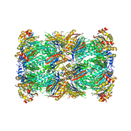
8W2F
DownloadVisualize
BU of 8w2f by Molmil
Plasmodium falciparum 20S proteasome bound to an inhibitor
Descriptor: (3S)-1-[(2-fluoroethoxy)acetyl]-N-{[(4P)-4-(6-methylpyridin-3-yl)-1,3-thiazol-2-yl]methyl}piperidine-3-carboxamide, Proteasome endopeptidase complex, Proteasome subunit alpha type, ...
Authors:Han, Y, Deng, X, Ray, S, Chen, Z, Phillips, M.
Deposit date:2024-02-20
Release date:2024-07-31
Last modified:2024-09-04
Method:ELECTRON MICROSCOPY (3.1 Å)
Cite:Identification of potent and reversible piperidine carboxamides that are species-selective orally active proteasome inhibitors to treat malaria.
Cell Chem Biol, 31, 2024
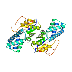
8WNH
DownloadVisualize
BU of 8wnh by Molmil
Crystal structure of cis-epoxysuccinate hydrolase from Rhodococcus opacus
Descriptor: Epoxide hydrolase, FORMIC ACID
Authors:Han, Y, Kong, X.D, Xu, J.H.
Deposit date:2023-10-05
Release date:2024-06-12
Last modified:2024-10-30
Method:X-RAY DIFFRACTION (1.773 Å)
Cite:Structural Insights of a cis -Epoxysuccinate Hydrolase Facilitate the Development of Robust Biocatalysts for the Production of l-(+)-Tartrate.
Biochemistry, 63, 2024
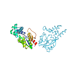
8WMT
DownloadVisualize
BU of 8wmt by Molmil
Crystal structure of cis-epoxysuccinic hydrolase from Bradyrhizobium mercantei
Descriptor: cis-epoxysuccinic hydrolase
Authors:Han, Y, Kong, X.D, Xu, J.H.
Deposit date:2023-10-04
Release date:2024-06-12
Last modified:2024-07-03
Method:X-RAY DIFFRACTION (1.72 Å)
Cite:Structural Insights of a cis -Epoxysuccinate Hydrolase Facilitate the Development of Robust Biocatalysts for the Production of l-(+)-Tartrate.
Biochemistry, 63, 2024
8WWS
DownloadVisualize
BU of 8wws by Molmil
Crystal structure of cis-epoxysuccinate hydrolase from Klebsiella oxytoca with L(+)-tartaric acid
Descriptor: (S)-2-haloacid dehalogenase, 1,2-ETHANEDIOL, CARBONATE ION, ...
Authors:Han, Y, Kong, X.D, Li, J, Xu, J.H.
Deposit date:2023-10-26
Release date:2024-06-12
Last modified:2024-07-03
Method:X-RAY DIFFRACTION (1.79 Å)
Cite:Structural Insights of a cis -Epoxysuccinate Hydrolase Facilitate the Development of Robust Biocatalysts for the Production of l-(+)-Tartrate.
Biochemistry, 63, 2024
6M1T
DownloadVisualize
BU of 6m1t by Molmil
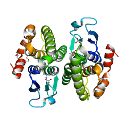
Bacterial beta class Sphingomonas chungbukensis Glutathione S-transferase
Descriptor: CYSTEINE, GLUTATHIONE, Glutathione S-transferase, ...
Authors:Han, Y.H, Chung, Y.J.
Deposit date:2020-02-26
Release date:2020-04-08
Last modified:2023-11-29
Method:X-RAY DIFFRACTION (1.9 Å)
Cite:Bacterial beta class Sphingomonas chungbukensis Glutathione S-transferase
To Be Published
4OAV
DownloadVisualize
BU of 4oav by Molmil
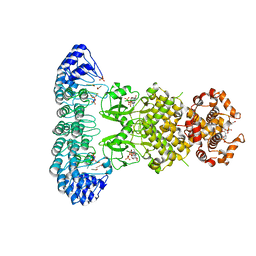
Complete human RNase L in complex with 2-5A (5'-ppp heptamer), AMPPCP and RNA substrate.
Descriptor: (2R,3S,4R,5R)-5-(2,4-dioxo-3,4-dihydropyrimidin-1(2H)-yl)-4-hydroxy-2-({[(S)-hydroxy{[(2R,3S,4S)-4-hydroxy-2-(hydroxymethyl)tetrahydrofuran-3-yl]oxy}phosphoryl]oxy}methyl)tetrahydrofuran-3-yl dihydrogen phosphate, MAGNESIUM ION, PHOSPHOMETHYLPHOSPHONIC ACID ADENYLATE ESTER, ...
Authors:Han, Y, Donovan, J, Rath, S, Whitney, G, Chitrakar, A, Korennykh, A.
Deposit date:2014-01-06
Release date:2014-03-12
Last modified:2023-09-20
Method:X-RAY DIFFRACTION (2.1 Å)
Cite:Structure of human RNase L reveals the basis for regulated RNA decay in the IFN response.
Science, 343, 2014
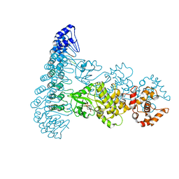
4OAU
DownloadVisualize
BU of 4oau by Molmil
Complete human RNase L in complex with biological activators.
Descriptor: 2-5A-dependent ribonuclease, ADENOSINE-5'-DIPHOSPHATE, MAGNESIUM ION, ...
Authors:Han, Y, Donovan, J, Rath, S, Whitney, G, Chitrakar, A, Korennykh, A.
Deposit date:2014-01-06
Release date:2014-03-12
Last modified:2024-02-28
Method:X-RAY DIFFRACTION (2.6 Å)
Cite:Structure of human RNase L reveals the basis for regulated RNA decay in the IFN response.
Science, 343, 2014

226707

PDB entries from 2024-10-30

PDB statisticsPDBj update infoContact PDBjnumon