Loading
PDBj
MenuPDBj@FacebookPDBj@TwitterPDBj@YouTubewwPDB FoundationwwPDB
RCSB PDBPDBeBMRBAdv. SearchSearch help
PDB: 160 results

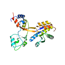
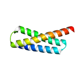
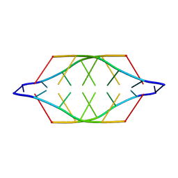
225D
DownloadVisualize
BU of 225d by Molmil
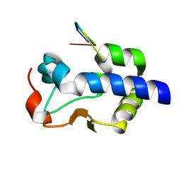
A TETRAMERIC DNA STRUCTURE WITH PROTONATED CYTOSINE:CYTOSINE BASE PAIRS
Descriptor: DNA (5'-D(*TP*CP*CP*CP*CP*C)-3')
Authors:Gehring, K, Leroy, J.L, Gueron, M.
Deposit date:1995-07-12
Release date:1995-10-15
Last modified:2024-05-22
Method:SOLUTION NMR
Cite:A tetrameric DNA structure with protonated cytosine.cytosine base pairs.
Nature, 363, 1993
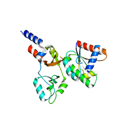
1D5G
DownloadVisualize
BU of 1d5g by Molmil
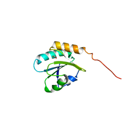
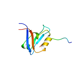
SOLUTION STRUCTURE OF THE PDZ2 DOMAIN FROM HUMAN PHOSPHATASE HPTP1E COMPLEXED WITH A PEPTIDE
Descriptor: HUMAN PHOSPHATASE HPTP1E, PEPTIDE FADSEADENEQVSAV
Authors:Kozlov, G, Gehring, K, Ekiel, I.
Deposit date:1999-10-07
Release date:2002-07-24
Last modified:2024-05-22
Method:SOLUTION NMR
Cite:Solution Structure of the PDZ2 Domain from Cytosolic Human Phosphatase hPTP1E Complexed with a Peptide Reveals Contribution of the beta2-beta3 Loop to PDZ Domain-Ligand Interactions
J.Mol.Biol., 320, 2002
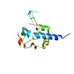
8SMO
DownloadVisualize
BU of 8smo by Molmil
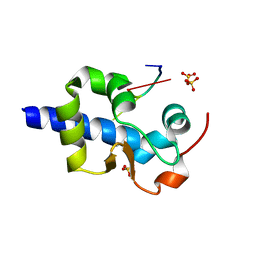
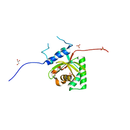
Crystal structure of the complex between truncated MLLE domain of PABPC1 and engineered superPAM2 peptide
Descriptor: Polyadenylate-binding protein 1, SULFATE ION, superPAM2
Authors:Kozlov, G, Gehring, K.
Deposit date:2023-04-26
Release date:2024-05-01
Method:X-RAY DIFFRACTION (3 Å)
Cite:Crystal structure of the complex between truncated MLLE domain of PABPC1 and engineered superPAM2 peptide
To Be Published
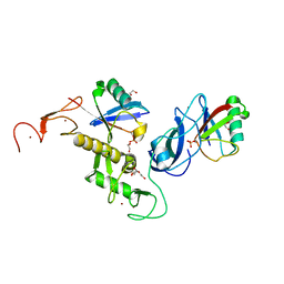
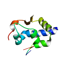
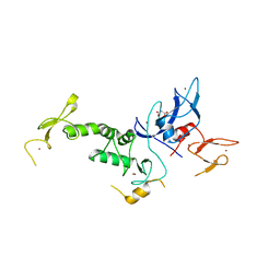
7US1
DownloadVisualize
BU of 7us1 by Molmil
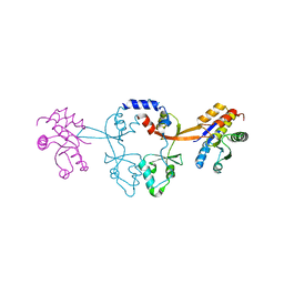
Structure of parkin (R0RB) bound to two phospho-ubiquitin molecules
Descriptor: 1,2-ETHANEDIOL, DI(HYDROXYETHYL)ETHER, E3 ubiquitin-protein ligase parkin, ...
Authors:Fakih, R, Sauve, V, Gehring, K.
Deposit date:2022-04-22
Release date:2022-06-22
Last modified:2023-10-18
Method:X-RAY DIFFRACTION (2.484 Å)
Cite:Structure of the second phosphoubiquitin-binding site in parkin.
J.Biol.Chem., 298, 2022
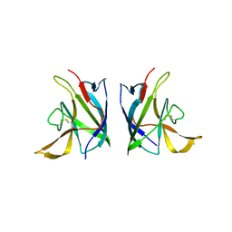
8GK6
DownloadVisualize
BU of 8gk6 by Molmil
Crystal structure of extracellular domain of CNNM4 from Echinococcus granulosus
Descriptor: 2-acetamido-2-deoxy-beta-D-glucopyranose, 2-acetamido-2-deoxy-beta-D-glucopyranose-(1-4)-2-acetamido-2-deoxy-beta-D-glucopyranose, Metal transporter CNNM4, ...
Authors:Shahsavan, A, Gehring, K.
Deposit date:2023-03-17
Release date:2023-12-13
Last modified:2024-02-07
Method:X-RAY DIFFRACTION (2.7 Å)
Cite:Dimerization of the CNNM extracellular domain.
Protein Sci., 33, 2024
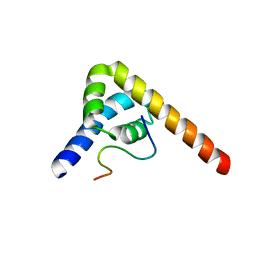
8G91
DownloadVisualize
BU of 8g91 by Molmil
LaM domain of human LARP1 in complex with Rp phosphorothioate isomer of AAAAA(SRA) RNA
Descriptor: GLYCEROL, Isoform 2 of La-related protein 1, RNA (5'-R(*AP*AP*AP*AP*AP*(SRA))-3')
Authors:Kozlov, G, Jiang, J, Gehring, K.
Deposit date:2023-02-20
Release date:2024-04-17
Method:X-RAY DIFFRACTION (1.2 Å)
Cite:Structural insights into RNA binding by the LaM domain of LARP1
To Be Published
8G90
DownloadVisualize
BU of 8g90 by Molmil
LaM domain of human LARP1 in complex with Sp phosphorothioate isomer of AAAAA(SRA) RNA
Descriptor: Isoform 2 of La-related protein 1, RNA (5'-R(*AP*AP*AP*AP*AP*(SRA))-3')
Authors:Kozlov, G, Jiang, J, Gehring, K.
Deposit date:2023-02-20
Release date:2024-04-17
Method:X-RAY DIFFRACTION (1.2 Å)
Cite:Structural insights into RNA binding by the LaM domain of LARP1
To Be Published
6PIK
DownloadVisualize
BU of 6pik by Molmil
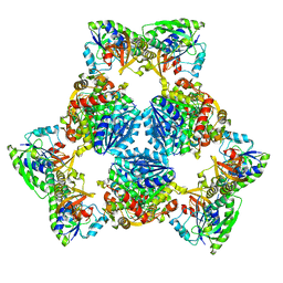
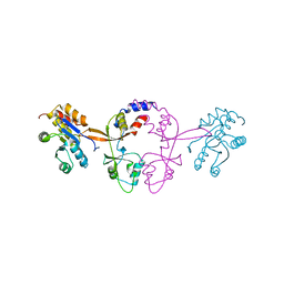
Tetrameric cryo-EM ArnA
Descriptor: Bifunctional polymyxin resistance protein ArnA, UDP-4-amino-4-deoxy-L-arabinose formyltransferase
Authors:Yang, M, Gehring, K.
Deposit date:2019-06-26
Release date:2019-07-31
Last modified:2024-03-20
Method:ELECTRON MICROSCOPY (7.8 Å)
Cite:Cryo-electron microscopy structures of ArnA, a key enzyme for polymyxin resistance, revealed unexpected oligomerizations and domain movements.
J.Struct.Biol., 208, 2019
6PIH
DownloadVisualize
BU of 6pih by Molmil
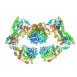
Hexameric ArnA cryo-EM structure
Descriptor: Bifunctional polymyxin resistance protein ArnA, UDP-4-amino-4-deoxy-L-arabinose formyltransferase
Authors:Yang, M, Gehring, K.
Deposit date:2019-06-26
Release date:2019-07-31
Last modified:2024-03-13
Method:ELECTRON MICROSCOPY (6.6 Å)
Cite:Cryo-electron microscopy structures of ArnA, a key enzyme for polymyxin resistance, revealed unexpected oligomerizations and domain movements.
J.Struct.Biol., 208, 2019
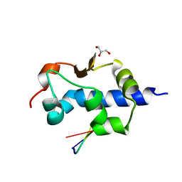
4NWY
DownloadVisualize
BU of 4nwy by Molmil
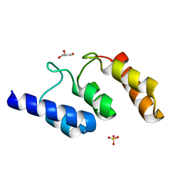
Crystal structure of the b' domain of human protein disulfide isomerase-like protein of the testis (PDILT)
Descriptor: FORMIC ACID, Protein disulfide-isomerase-like protein of the testis
Authors:Bastos-Aristizabal, S, Kozlov, G, Gehring, K.
Deposit date:2013-12-07
Release date:2014-04-02
Last modified:2023-09-20
Method:X-RAY DIFFRACTION (2 Å)
Cite:Structure of the substrate-binding b' domain of the Protein disulfide isomerase-like protein of the testis.
Sci Rep, 4, 2014
8CT8
DownloadVisualize
BU of 8ct8 by Molmil
Crystal structure of Drosophila melanogaster PRL/CBS-pair domain complex
Descriptor: IODIDE ION, PRL-1 phosphatase, Unextended protein
Authors:Fakih, R, Goldstein, R.H, Kozlov, G, Gehring, K.
Deposit date:2022-05-13
Release date:2023-03-01
Last modified:2024-05-22
Method:X-RAY DIFFRACTION (2.5 Å)
Cite:Burst kinetics and CNNM binding are evolutionarily conserved properties of phosphatases of regenerating liver.
J.Biol.Chem., 299, 2023
8F6D
DownloadVisualize
BU of 8f6d by Molmil
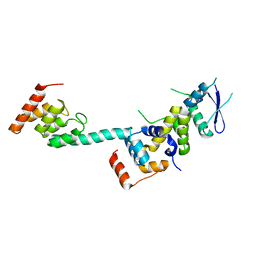
Crystal structure of the CNNM2 CBS-pair domain in complex with ARL15
Descriptor: ADP-ribosylation factor-like protein 15, Metal transporter CNNM2
Authors:Kozlov, G, Mahbub, L, Gehring, K.
Deposit date:2022-11-16
Release date:2023-07-12
Last modified:2024-01-24
Method:X-RAY DIFFRACTION (3.2 Å)
Cite:Structural insights into regulation of CNNM-TRPM7 divalent cation uptake by the small GTPase ARL15.
Elife, 12, 2023
8EY7
DownloadVisualize
BU of 8ey7 by Molmil
LaM domain of human LARP1 in complex with AAAGAA RNA
Descriptor: Isoform 2 of La-related protein 1, RNA (5'-R(*AP*AP*AP*GP*AP*A)-3')
Authors:Kozlov, G, Gehring, K.
Deposit date:2022-10-26
Release date:2023-11-01
Method:X-RAY DIFFRACTION (1.35 Å)
Cite:Structural insights into poly(A) tail stabilization via 3'-end guanylation
To Be Published
8EY8
DownloadVisualize
BU of 8ey8 by Molmil
LaM domain of human LARP1 in complex with AAAAGA RNA
Descriptor: Isoform 2 of La-related protein 1, RNA (5'-R(*AP*AP*AP*AP*GP*A)-3')
Authors:Kozlov, G, Gehring, K.
Deposit date:2022-10-26
Release date:2023-11-01
Method:X-RAY DIFFRACTION (1.3 Å)
Cite:Structural insights into poly(A) tail stabilization via 3'-end guanylation
To Be Published
8EY6
DownloadVisualize
BU of 8ey6 by Molmil
LaM domain of human LARP1 in complex with AAAAAG RNA
Descriptor: Isoform 2 of La-related protein 1, RNA (5'-R(*AP*AP*AP*AP*AP*G)-3'), SULFATE ION
Authors:Kozlov, G, Gehring, K.
Deposit date:2022-10-26
Release date:2023-11-01
Method:X-RAY DIFFRACTION (1.63 Å)
Cite:Structural insights into poly(A) tail stabilization via 3'-end guanylation
To Be Published
6WUS
DownloadVisualize
BU of 6wus by Molmil
Crystal structure of PRL-1 phosphatase C104D mutant in complex with the Bateman domain of CNNM2 magnesium transporter
Descriptor: Metal transporter CNNM2, Protein tyrosine phosphatase type IVA 1
Authors:Kozlov, G, Gehring, K.
Deposit date:2020-05-05
Release date:2020-07-01
Last modified:2023-10-18
Method:X-RAY DIFFRACTION (2.758 Å)
Cite:Crystal structure of PRL phosphatase C104D mutant in complex with the Bateman domain of CNNM magnesium transporter
J.Biol.Chem., 2020
6WUR
DownloadVisualize
BU of 6wur by Molmil
Crystal structure of PRL-2 phosphatase C101D mutant in complex with the Bateman domain of CNNM3 magnesium transporter
Descriptor: Metal transporter CNNM3, Protein tyrosine phosphatase type IVA 2, SODIUM ION
Authors:Kozlov, G, Gehring, K.
Deposit date:2020-05-05
Release date:2020-07-01
Last modified:2023-10-18
Method:X-RAY DIFFRACTION (2.882 Å)
Cite:Crystal structure of PRL phosphatase C104D mutant in complex with the Bateman domain of CNNM magnesium transporter
J.Biol.Chem., 2020
5K35
DownloadVisualize
BU of 5k35 by Molmil
Structure of the Legionella effector, AnkB, in complex with human Skp1
Descriptor: Ankyrin-repeat protein B, S-phase kinase-associated protein 1
Authors:Wong, K, Kozlov, G, Gehring, K, Montreal-Kingston Bacterial Structural Genomics Initiative (BSGI)
Deposit date:2016-05-19
Release date:2017-01-25
Last modified:2024-03-06
Method:X-RAY DIFFRACTION (2.85 Å)
Cite:Structural Mimicry by a Bacterial F Box Effector Hijacks the Host Ubiquitin-Proteasome System.
Structure, 25, 2017
5K34
DownloadVisualize
BU of 5k34 by Molmil
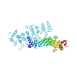
Structure of the ankyrin domain of AnkB from Legionella Pneumophila
Descriptor: Ankyrin-repeat protein B, GLYCEROL, SULFATE ION
Authors:Wong, K, Kozlov, G, Gehring, K, Montreal-Kingston Bacterial Structural Genomics Initiative (BSGI)
Deposit date:2016-05-19
Release date:2017-01-25
Last modified:2024-03-06
Method:X-RAY DIFFRACTION (1.15 Å)
Cite:Structural Mimicry by a Bacterial F Box Effector Hijacks the Host Ubiquitin-Proteasome System.
Structure, 25, 2017
5KDG
DownloadVisualize
BU of 5kdg by Molmil
Crystal Structure of Salmonella Typhimurium Effector GtgE
Descriptor: GLYCEROL, Gifsy-2 prophage protein, SULFATE ION
Authors:Kozlov, G, Xu, C, Wong, K, Gehring, K, Cygler, M, Montreal-Kingston Bacterial Structural Genomics Initiative (BSGI)
Deposit date:2016-06-08
Release date:2016-11-16
Last modified:2023-09-27
Method:X-RAY DIFFRACTION (1.73 Å)
Cite:Crystal Structure of the Salmonella Typhimurium Effector GtgE.
PLoS ONE, 11, 2016
5VRQ
DownloadVisualize
BU of 5vrq by Molmil
Crystal structure of Legionella pneumophila effector AnkC
Descriptor: Ankyrin repeat-containing protein
Authors:Kozlov, G, Wong, K, Wang, W, Skubak, P, Munoz-Escobar, J, Liu, Y, Pannu, N.S, Gehring, K, Montreal-Kingston Bacterial Structural Genomics Initiative (BSGI)
Deposit date:2017-05-11
Release date:2017-11-29
Last modified:2024-03-13
Method:X-RAY DIFFRACTION (3.205 Å)
Cite:Ankyrin repeats as a dimerization module.
Biochem. Biophys. Res. Commun., 495, 2018
5WD8
DownloadVisualize
BU of 5wd8 by Molmil
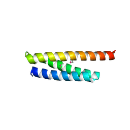
Crystal structure of Legionella pneumophila effector lpg2328
Descriptor: Lem22
Authors:Kozlov, G, Wong, K, Gehring, K, Montreal-Kingston Bacterial Structural Genomics Initiative (BSGI)
Deposit date:2017-07-04
Release date:2017-11-29
Last modified:2020-01-08
Method:X-RAY DIFFRACTION (1.944 Å)
Cite:Crystal structure of the Legionella effector Lem22.
Proteins, 86, 2018
5WD9
DownloadVisualize
BU of 5wd9 by Molmil
Crystal structure of Legionella pneumophila effector lpg2328
Descriptor: Lem22
Authors:Kozlov, G, Wong, K, Gehring, K, Montreal-Kingston Bacterial Structural Genomics Initiative (BSGI)
Deposit date:2017-07-04
Release date:2017-11-29
Last modified:2024-04-03
Method:X-RAY DIFFRACTION (1.4 Å)
Cite:Crystal structure of the Legionella effector Lem22.
Proteins, 86, 2018
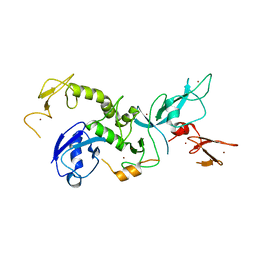
4K7D
DownloadVisualize
BU of 4k7d by Molmil
Crystal Structure of Parkin C-terminal RING domains
Descriptor: CHLORIDE ION, E3 ubiquitin-protein ligase parkin, MALONATE ION, ...
Authors:Sauve, V, Trempe, J.-F, Menade, M, Gehring, K.
Deposit date:2013-04-17
Release date:2013-05-15
Last modified:2024-02-28
Method:X-RAY DIFFRACTION (2.8 Å)
Cite:Structure of parkin reveals mechanisms for ubiquitin ligase activation.
Science, 340, 2013
4K95
DownloadVisualize
BU of 4k95 by Molmil
Crystal Structure of Parkin
Descriptor: E3 ubiquitin-protein ligase parkin, ZINC ION
Authors:Seirafi, M, Menade, M, Sauve, V, Kozlov, G, Trempe, J.-F, Nagar, B, Gehring, K.
Deposit date:2013-04-19
Release date:2013-05-15
Last modified:2023-09-20
Method:X-RAY DIFFRACTION (6.499 Å)
Cite:Structure of parkin reveals mechanisms for ubiquitin ligase activation.
Science, 340, 2013

220472

PDB entries from 2024-05-29

PDB statisticsPDBj update infoContact PDBjnumon