Loading
PDBj
MenuPDBj@FacebookPDBj@TwitterPDBj@YouTubewwPDB FoundationwwPDB
RCSB PDBPDBeBMRBAdv. SearchSearch help
PDB: 180 results

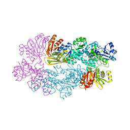
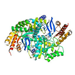
3BV0
DownloadVisualize
BU of 3bv0 by Molmil
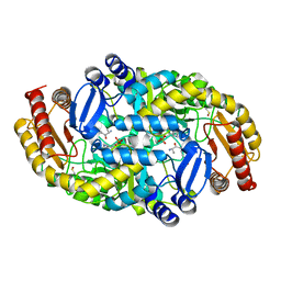
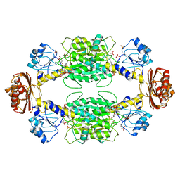
Crystal Structure of PLP Bound 7,8-Diaminopelargonic Acid Synthase in Mycobacterium Tuberculosis
Descriptor: Adenosylmethionine-8-amino-7-oxononanoate aminotransferase, PYRIDOXAL-5'-PHOSPHATE
Authors:Dey, S, Sacchettini, J.C.
Deposit date:2008-01-04
Release date:2008-02-05
Last modified:2023-08-30
Method:X-RAY DIFFRACTION (2.21 Å)
Cite:Structural characterization of the Mycobacterium tuberculosis biotin biosynthesis enzymes 7,8-diaminopelargonic acid synthase and dethiobiotin synthetase .
Biochemistry, 49, 2010
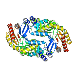
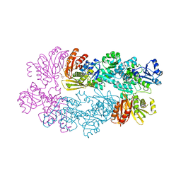
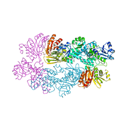
5ZZM
DownloadVisualize
BU of 5zzm by Molmil
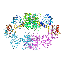
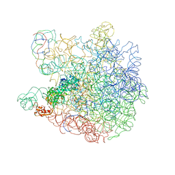
E. coli 50S subunit bound HflX protein in presence of ATP (AMP-PNP) and GTP (GMP-PNP) analogs.
Descriptor: 23S rRNA, 5S rRNA, GTPase HflX
Authors:Dey, S.
Deposit date:2018-06-03
Release date:2018-06-27
Last modified:2024-03-27
Method:ELECTRON MICROSCOPY (8.1 Å)
Cite:The universally conserved GTPase HflX is an RNA helicase that restores heat-damagedEscherichia coliribosomes.
J. Cell Biol., 217, 2018
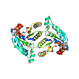
6CJD
DownloadVisualize
BU of 6cjd by Molmil
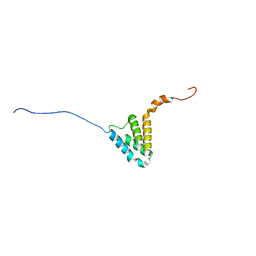
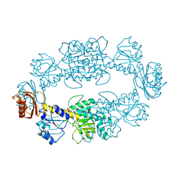
NMR Structure of Salmonella Type III Secretion system protein OrgC
Descriptor: Putative cytoplasmic protein
Authors:DEY, S, DE GUZMAN, R.N.
Deposit date:2018-02-26
Release date:2018-08-01
Last modified:2023-06-14
Method:SOLUTION NMR
Cite:A protein secreted by theSalmonellatype III secretion system controls needle filament assembly.
Elife, 7, 2018
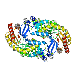
8ABY
DownloadVisualize
BU of 8aby by Molmil
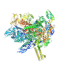
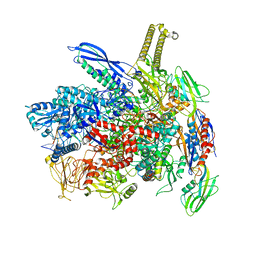
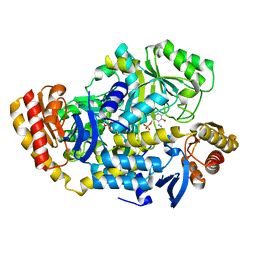
RNA polymerase bound to purified in vitro transcribed regulatory RNA putL - pause prone, closed clamp state
Descriptor: DNA-directed RNA polymerase subunit alpha, DNA-directed RNA polymerase subunit beta, DNA-directed RNA polymerase subunit beta', ...
Authors:Dey, S, Weixlbaumer, A.
Deposit date:2022-07-05
Release date:2022-10-19
Last modified:2022-11-02
Method:ELECTRON MICROSCOPY (3.7 Å)
Cite:Structural insights into RNA-mediated transcription regulation in bacteria.
Mol.Cell, 82, 2022
8AD1
DownloadVisualize
BU of 8ad1 by Molmil
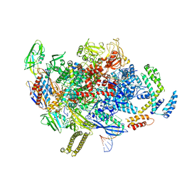
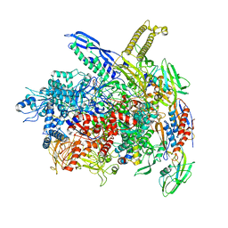
RNA polymerase at U-rich pause bound to RNA putL triple mutant - pause prone, closed clamp state
Descriptor: DNA-directed RNA polymerase subunit alpha, DNA-directed RNA polymerase subunit beta, DNA-directed RNA polymerase subunit beta', ...
Authors:Dey, S, Weixlbaumer, A.
Deposit date:2022-07-07
Release date:2022-10-19
Last modified:2022-11-02
Method:ELECTRON MICROSCOPY (4.1 Å)
Cite:Structural insights into RNA-mediated transcription regulation in bacteria.
Mol.Cell, 82, 2022
8ACP
DownloadVisualize
BU of 8acp by Molmil
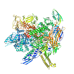
RNA polymerase at U-rich pause bound to regulatory RNA putL - inactive, open clamp state
Descriptor: DNA-directed RNA polymerase subunit alpha, DNA-directed RNA polymerase subunit beta, DNA-directed RNA polymerase subunit beta', ...
Authors:Dey, S, Weixlbaumer, A.
Deposit date:2022-07-06
Release date:2022-10-19
Last modified:2022-11-02
Method:ELECTRON MICROSCOPY (4.5 Å)
Cite:Structural insights into RNA-mediated transcription regulation in bacteria.
Mol.Cell, 82, 2022
8ABZ
DownloadVisualize
BU of 8abz by Molmil
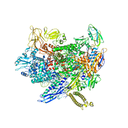
RNA polymerase at U-rich pause bound to non-regulatory RNA - pause prone, closed clamp state
Descriptor: DNA-directed RNA polymerase subunit alpha, DNA-directed RNA polymerase subunit beta, DNA-directed RNA polymerase subunit beta', ...
Authors:Dey, S, Weixlbaumer, A.
Deposit date:2022-07-05
Release date:2022-10-19
Last modified:2022-11-02
Method:ELECTRON MICROSCOPY (3.4 Å)
Cite:Structural insights into RNA-mediated transcription regulation in bacteria.
Mol.Cell, 82, 2022
8AC2
DownloadVisualize
BU of 8ac2 by Molmil
RNA polymerase- post-terminated, open clamp state
Descriptor: DNA Non-template strand, DNA Template strand, DNA-directed RNA polymerase subunit alpha, ...
Authors:Dey, S, Weixlbaumer, A.
Deposit date:2022-07-05
Release date:2022-10-19
Last modified:2022-11-02
Method:ELECTRON MICROSCOPY (3.7 Å)
Cite:Structural insights into RNA-mediated transcription regulation in bacteria.
Mol.Cell, 82, 2022
8AC1
DownloadVisualize
BU of 8ac1 by Molmil
RNA polymerase at U-rich pause bound to non-regulatory RNA - inactive, open clamp state
Descriptor: DNA Non-template strand, DNA Template strand, DNA-directed RNA polymerase subunit alpha, ...
Authors:Dey, S, Weixlbaumer, A.
Deposit date:2022-07-05
Release date:2022-10-26
Last modified:2022-11-02
Method:ELECTRON MICROSCOPY (4.06 Å)
Cite:Structural insights into RNA-mediated transcription regulation in bacteria.
Mol.Cell, 82, 2022
4QHT
DownloadVisualize
BU of 4qht by Molmil
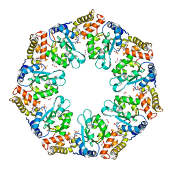
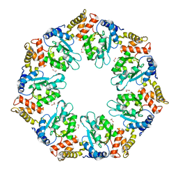
Crystal structure of AAA+/ sigma 54 activator domain of the flagellar regulatory protein FlrC from Vibrio cholerae in ATP analog bound state
Descriptor: 1,2-ETHANEDIOL, Flagellar regulatory protein C, MAGNESIUM ION, ...
Authors:Dey, S, Biswas, M, Sen, U, Dasgupta, J.
Deposit date:2014-05-29
Release date:2014-07-16
Last modified:2024-03-20
Method:X-RAY DIFFRACTION (2.559 Å)
Cite:Unique ATPase site architecture triggers cis-mediated synchronized ATP binding in heptameric AAA+-ATPase domain of flagellar regulatory protein FlrC
J.Biol.Chem., 290, 2015
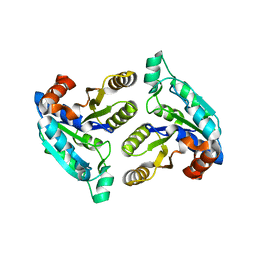
3FPA
DownloadVisualize
BU of 3fpa by Molmil
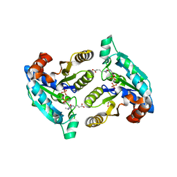
Crystal structure of Mycobacterium tuberculosis dethiobiotin synthetase complexed with dethiobiotin and phosphate
Descriptor: 6-(5-METHYL-2-OXO-IMIDAZOLIDIN-4-YL)-HEXANOIC ACID, Dethiobiotin synthetase, MAGNESIUM ION, ...
Authors:Dey, S, Sacchettini, J.C.
Deposit date:2009-01-04
Release date:2009-06-30
Last modified:2023-09-06
Method:X-RAY DIFFRACTION (2.3 Å)
Cite:Structural characterization of the Mycobacterium tuberculosis biotin biosynthesis enzymes 7,8-diaminopelargonic acid synthase and dethiobiotin synthetase.
Biochemistry, 49, 2010
4QHS
DownloadVisualize
BU of 4qhs by Molmil
Crystal structure of AAA+sigma 54 activator domain of the flagellar regulatory protein FlrC of Vibrio cholerae in nucleotide free state
Descriptor: 1,2-ETHANEDIOL, Flagellar regulatory protein C
Authors:Dey, S, Biswas, M, Sen, U, Dasgupta, J.
Deposit date:2014-05-29
Release date:2014-07-16
Last modified:2023-11-08
Method:X-RAY DIFFRACTION (2.3 Å)
Cite:Unique ATPase site architecture triggers cis-mediated synchronized ATP binding in heptameric AAA+-ATPase domain of flagellar regulatory protein FlrC
J.Biol.Chem., 290, 2015
3FMI
DownloadVisualize
BU of 3fmi by Molmil
Crystal structure of Mycobacterium tuberculosis dethiobiotin synthetase complexed with 7-Keto 8-aminopelargonic acid
Descriptor: 7-KETO-8-AMINOPELARGONIC ACID, Dethiobiotin synthetase, SULFATE ION
Authors:Dey, S, Sacchettini, J.C.
Deposit date:2008-12-22
Release date:2009-06-30
Last modified:2023-09-06
Method:X-RAY DIFFRACTION (2.18 Å)
Cite:Structural characterization of the Mycobacterium tuberculosis biotin biosynthesis enzymes 7,8-diaminopelargonic acid synthase and dethiobiotin synthetase .
Biochemistry, 49, 2010
3DU4
DownloadVisualize
BU of 3du4 by Molmil
Crystal structure of 7-keto-8-aminopelargonic acid bound 7,8-diaminopelargonic acid synthase in bacillus subtilis
Descriptor: 7-KETO-8-AMINOPELARGONIC ACID, Adenosylmethionine-8-amino-7-oxononanoate aminotransferase, PYRIDOXAL-5'-PHOSPHATE
Authors:Dey, S, Sacchettini, J.C.
Deposit date:2008-07-16
Release date:2009-06-23
Last modified:2023-08-30
Method:X-RAY DIFFRACTION (2.2 Å)
Cite:Structural characterization of the Mycobacterium tuberculosis biotin biosynthesis enzymes 7,8-diaminopelargonic acid synthase and dethiobiotin synthetase .
Biochemistry, 49, 2010
1YGY
DownloadVisualize
BU of 1ygy by Molmil
Crystal Structure of D-3-Phosphoglycerate dehydrogenase From Mycobacterium tuberculosis
Descriptor: D-3-phosphoglycerate dehydrogenase, L(+)-TARTARIC ACID
Authors:Dey, S, Grant, G.A, Sacchettini, J.C, TB Structural Genomics Consortium (TBSGC)
Deposit date:2005-01-05
Release date:2005-01-25
Last modified:2019-07-10
Method:X-RAY DIFFRACTION (2.3 Å)
Cite:Crystal Structure of Mycobacterium tuberculosis D-3-Phosphoglycerate Dehydrogenase: EXTREME ASYMMETRY IN A TETRAMER OF IDENTICAL SUBUNITS
J.Biol.Chem., 280, 2005
3DRD
DownloadVisualize
BU of 3drd by Molmil
Crystal Structure of 7,8 Diaminopelargonic Acid Synthase Apoenzyme in Bacillus subtilis
Descriptor: Adenosylmethionine-8-amino-7-oxononanoate aminotransferase
Authors:Dey, S, Sacchettini, J.C.
Deposit date:2008-07-11
Release date:2009-06-23
Last modified:2023-08-30
Method:X-RAY DIFFRACTION (2.17 Å)
Cite:Structural characterization of the Mycobacterium tuberculosis biotin biosynthesis enzymes 7,8-diaminopelargonic acid synthase and dethiobiotin synthetase .
Biochemistry, 49, 2010
3FMF
DownloadVisualize
BU of 3fmf by Molmil
Crystal structure of Mycobacterium tuberculosis dethiobiotin synthetase complexed with 7,8 diaminopelargonic acid carbamate
Descriptor: 7-(CARBOXYAMINO)-8-AMINO-NONANOIC ACID, Dethiobiotin synthetase
Authors:Dey, S, Sacchettini, J.C.
Deposit date:2008-12-21
Release date:2009-06-30
Last modified:2023-09-06
Method:X-RAY DIFFRACTION (2.05 Å)
Cite:Structural characterization of the Mycobacterium tuberculosis biotin biosynthesis enzymes 7,8-diaminopelargonic acid synthase and dethiobiotin synthetase .
Biochemistry, 49, 2010
3DOD
DownloadVisualize
BU of 3dod by Molmil
Crystal Structure of PLP Bound 7,8-Diaminopelargonic Acid Synthase in Bacillus subtilis
Descriptor: Adenosylmethionine-8-amino-7-oxononanoate aminotransferase, PYRIDOXAL-5'-PHOSPHATE
Authors:Dey, S, Sacchettini, J.C.
Deposit date:2008-07-03
Release date:2009-06-23
Last modified:2023-08-30
Method:X-RAY DIFFRACTION (1.9 Å)
Cite:Structural characterization of the Mycobacterium tuberculosis biotin biosynthesis enzymes 7,8-diaminopelargonic acid synthase and dethiobiotin synthetase .
Biochemistry, 49, 2010
3DC2
DownloadVisualize
BU of 3dc2 by Molmil
Crystal structure of serine bound D-3-phosphoglycerate dehydrogenase from Mycobacterium tuberculosis
Descriptor: D-3-phosphoglycerate dehydrogenase, L(+)-TARTARIC ACID, SERINE
Authors:Dey, S, Sacchettini, J.C.
Deposit date:2008-06-03
Release date:2008-07-22
Last modified:2023-11-01
Method:X-RAY DIFFRACTION (2.7 Å)
Cite:Structural analysis of substrate and effector binding in Mycobacterium tuberculosis D-3-phosphoglycerate dehydrogenase
Biochemistry, 47, 2008
3FGN
DownloadVisualize
BU of 3fgn by Molmil
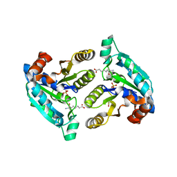
Crystal structure of dethiobiotin synthetase in Mycobacterium tuberculosis
Descriptor: Dethiobiotin synthetase
Authors:Dey, S, Sacchettini, J.C.
Deposit date:2008-12-08
Release date:2009-06-30
Last modified:2024-02-21
Method:X-RAY DIFFRACTION (1.85 Å)
Cite:Structural characterization of the Mycobacterium tuberculosis biotin biosynthesis enzymes 7,8-diaminopelargonic acid synthase and dethiobiotin synthetase .
Biochemistry, 49, 2010
3DDN
DownloadVisualize
BU of 3ddn by Molmil
Crystal structure of hydroxypyruvic acid phosphate bound D-3-phosphoglycerate dehydrogenase in mycobacterium tuberculosis
Descriptor: 2-oxo-3-(phosphonooxy)propanoic acid, D-3-phosphoglycerate dehydrogenase, L(+)-TARTARIC ACID
Authors:Dey, S, Sacchettini, J.C.
Deposit date:2008-06-05
Release date:2008-07-22
Last modified:2023-11-01
Method:X-RAY DIFFRACTION (2.4 Å)
Cite:Structural analysis of substrate and effector binding in Mycobacterium tuberculosis D-3-phosphoglycerate dehydrogenase
Biochemistry, 47, 2008
3LV2
DownloadVisualize
BU of 3lv2 by Molmil
Crystal structure of Mycobacterium tuberculosis 7,8-diaminopelargonic acid synthase in complex with substrate analog sinefungin
Descriptor: Adenosylmethionine-8-amino-7-oxononanoate aminotransferase, PYRIDOXAL-5'-PHOSPHATE, SINEFUNGIN
Authors:Dey, S, Sacchettini, J.C.
Deposit date:2010-02-19
Release date:2010-08-25
Last modified:2023-09-06
Method:X-RAY DIFFRACTION (2.18 Å)
Cite:Structural characterization of the Mycobacterium tuberculosis biotin biosynthesis enzymes 7,8-diaminopelargonic acid synthase and dethiobiotin synthetase .
Biochemistry, 49, 2010
2P9E
DownloadVisualize
BU of 2p9e by Molmil
Crystal Structure of G336V mutant of E.coli phosphoglycerate dehydrogenase
Descriptor: 1,4-DIHYDRONICOTINAMIDE ADENINE DINUCLEOTIDE, CITRIC ACID, D-3-phosphoglycerate dehydrogenase, ...
Authors:Dey, S, Sacchettini, J.C.
Deposit date:2007-03-25
Release date:2007-04-24
Last modified:2023-08-30
Method:X-RAY DIFFRACTION (2.6 Å)
Cite:The Effect of Hinge Mutations on Effector Binding and Domain Rotation in Escherichia coli D-3-Phosphoglycerate Dehydrogenase
J.Biol.Chem., 282, 2007
2P9C
DownloadVisualize
BU of 2p9c by Molmil
Crystal structure of serine bound G336V mutant of E.coli phosphoglycerate dehydrogenase
Descriptor: 1,4-DIHYDRONICOTINAMIDE ADENINE DINUCLEOTIDE, D-3-phosphoglycerate dehydrogenase, SERINE
Authors:Dey, S, Sacchettini, J.C.
Deposit date:2007-03-24
Release date:2007-04-24
Last modified:2023-08-30
Method:X-RAY DIFFRACTION (2.46 Å)
Cite:The Effect of Hinge Mutations on Effector Binding and Domain Rotation in Escherichia coli D-3-Phosphoglycerate Dehydrogenase
J.Biol.Chem., 282, 2007
2PA3
DownloadVisualize
BU of 2pa3 by Molmil
crystal structure of serine bound G336V mutant of E.coli phosphoglycerate dehydrogenase
Descriptor: 1,4-DIHYDRONICOTINAMIDE ADENINE DINUCLEOTIDE, D-3-phosphoglycerate dehydrogenase, SERINE
Authors:Dey, S, Sacchettini, J.C.
Deposit date:2007-03-27
Release date:2007-04-24
Last modified:2023-08-30
Method:X-RAY DIFFRACTION (2.74 Å)
Cite:The Effect of Hinge Mutations on Effector Binding and Domain Rotation in Escherichia coli D-3-Phosphoglycerate Dehydrogenase.
J.Biol.Chem., 282, 2007

219140

PDB entries from 2024-05-01

PDB statisticsPDBj update infoContact PDBjnumon