Loading
PDBj
MenuPDBj@FacebookPDBj@TwitterPDBj@YouTubewwPDB FoundationwwPDB
RCSB PDBPDBeBMRBAdv. SearchSearch help
Search by PDB author
2QHO
DownloadVisualize
BU of 2qho by Molmil
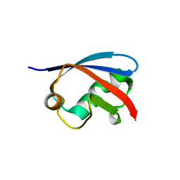
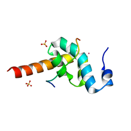
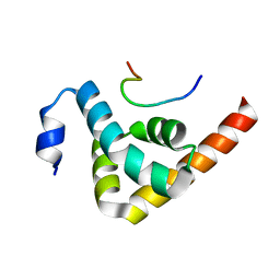
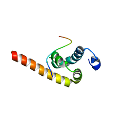
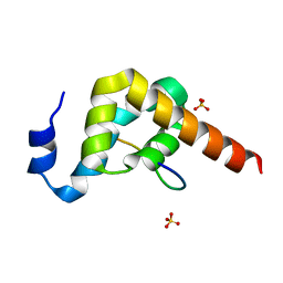
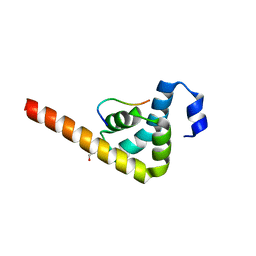
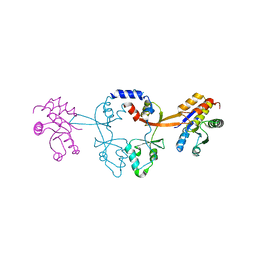
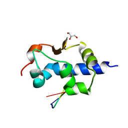
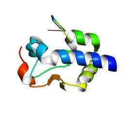
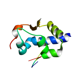
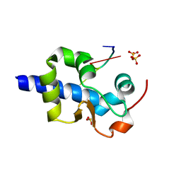
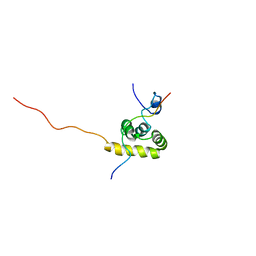
Crystal structure of the UBA domain from EDD ubiquitin ligase in complex with ubiquitin
Descriptor: E3 ubiquitin-protein ligase EDD1, Ubiquitin
Authors:Kozlov, G, Gehring, K.
Deposit date:2007-07-02
Release date:2007-09-25
Last modified:2023-08-30
Method:X-RAY DIFFRACTION (1.85 Å)
Cite:Structural basis of ubiquitin recognition by the ubiquitin-associated (UBA) domain of the ubiquitin ligase EDD.
J.Biol.Chem., 282, 2007
1L1P
DownloadVisualize
BU of 1l1p by Molmil
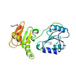
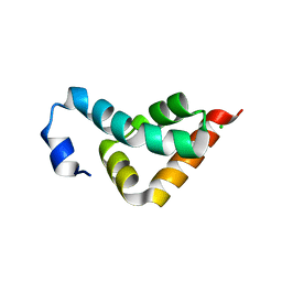
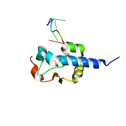
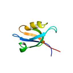
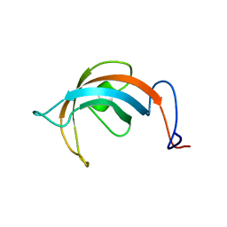
Solution Structure of the PPIase Domain from E. coli Trigger Factor
Descriptor: trigger factor
Authors:Kozlov, G, Trempe, J.-F, Perreault, A, Wong, M, Denisov, A, Ghandi, S, Gehring, K, Ekiel, I, Montreal-Kingston Bacterial Structural Genomics Initiative (BSGI)
Deposit date:2002-02-19
Release date:2003-06-24
Last modified:2022-02-23
Method:SOLUTION NMR
Cite:Solution Structure of the Closed Form of a Peptidyl-Prolyl Isomerase Reveals the Mechanism of Protein Folding
To be Published
4EEW
DownloadVisualize
BU of 4eew by Molmil
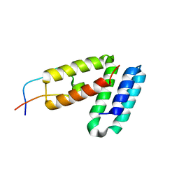
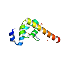
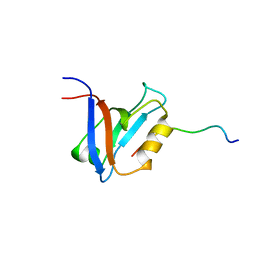
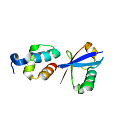
Crystal structure of the Ubl domain of BAG6
Descriptor: Large proline-rich protein BAG6
Authors:Kozlov, G, LePage, K, Vinaik, R, Gehring, K.
Deposit date:2012-03-28
Release date:2012-04-11
Last modified:2023-09-13
Method:X-RAY DIFFRACTION (1.3 Å)
Cite:Crystal structure of the Ubl domain of BAG6
To be Published
5V8Z
DownloadVisualize
BU of 5v8z by Molmil
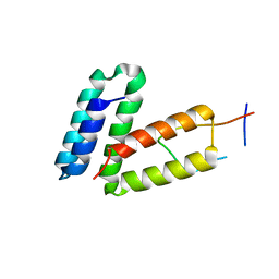
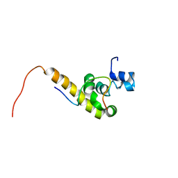
Crystal structure of ERp29 D-domain in complex with the P-domain of calmegin
Descriptor: Calmegin, Endoplasmic reticulum resident protein 29
Authors:Kozlov, G, Munoz-Escobar, J, Gehring, K.
Deposit date:2017-03-22
Release date:2017-06-21
Last modified:2023-10-04
Method:X-RAY DIFFRACTION (2.105 Å)
Cite:Mapping the ER Interactome: The P Domains of Calnexin and Calreticulin as Plurivalent Adapters for Foldases and Chaperones.
Structure, 25, 2017
5V90
DownloadVisualize
BU of 5v90 by Molmil
Crystal structure of ERp29 D-domain in complex with the P-domain of calreticulin
Descriptor: Calreticulin, Endoplasmic reticulum resident protein 29, GLYCEROL
Authors:Kozlov, G, Munoz-Escobar, J, Gehring, K.
Deposit date:2017-03-22
Release date:2017-06-21
Last modified:2023-10-04
Method:X-RAY DIFFRACTION (3.255 Å)
Cite:Mapping the ER Interactome: The P Domains of Calnexin and Calreticulin as Plurivalent Adapters for Foldases and Chaperones.
Structure, 25, 2017
3EC3
DownloadVisualize
BU of 3ec3 by Molmil
Crystal structure of the bb fragment of ERp72
Descriptor: Protein disulfide-isomerase A4
Authors:Kozlov, G, Gehring, K.
Deposit date:2008-08-28
Release date:2009-04-14
Last modified:2021-10-20
Method:X-RAY DIFFRACTION (1.92 Å)
Cite:Structure of the Noncatalytic Domains and Global Fold of the Protein Disulfide Isomerase ERp72.
Structure, 17, 2009
3KTR
DownloadVisualize
BU of 3ktr by Molmil
Structural basis of ataxin-2 recognition by poly(A)-binding protein
Descriptor: Ataxin-2, CADMIUM ION, Polyadenylate-binding protein 1, ...
Authors:Kozlov, G, Gehring, K.
Deposit date:2009-11-25
Release date:2010-02-23
Last modified:2023-09-06
Method:X-RAY DIFFRACTION (1.7 Å)
Cite:Structural basis of binding of P-body-associated proteins GW182 and ataxin-2 by the Mlle domain of poly(A)-binding protein.
J.Biol.Chem., 285, 2010
3KTP
DownloadVisualize
BU of 3ktp by Molmil
Structural basis of GW182 recognition by poly(A)-binding protein
Descriptor: Polyadenylate-binding protein 1, Trinucleotide repeat-containing gene 6C protein
Authors:Kozlov, G, Gehring, K.
Deposit date:2009-11-25
Release date:2010-02-23
Last modified:2023-09-06
Method:X-RAY DIFFRACTION (1.5 Å)
Cite:Structural basis of binding of P-body-associated proteins GW182 and ataxin-2 by the Mlle domain of poly(A)-binding protein.
J.Biol.Chem., 285, 2010
3KUT
DownloadVisualize
BU of 3kut by Molmil
Crystal structure of the MLLE domain of poly(A)-binding protein in complex with the binding region of Paip2
Descriptor: CHLORIDE ION, PAIP2 protein, Polyadenylate-binding protein 1
Authors:Kozlov, G, Gehring, K.
Deposit date:2009-11-27
Release date:2010-02-09
Last modified:2023-09-06
Method:X-RAY DIFFRACTION (1.5 Å)
Cite:Molecular Determinants of PAM2 Recognition by the MLLE Domain of Poly(A)-Binding Protein.
J.Mol.Biol., 397, 2010
3KUJ
DownloadVisualize
BU of 3kuj by Molmil
Crystal structure of C-terminal domain of PABPC1 in complex with binding region of eRF3a
Descriptor: GSPT1 protein, Polyadenylate-binding protein 1, SULFATE ION
Authors:Kozlov, G, Gehring, K.
Deposit date:2009-11-27
Release date:2010-05-12
Last modified:2023-09-06
Method:X-RAY DIFFRACTION (1.4 Å)
Cite:Molecular basis of eRF3 recognition by the MLLE domain of poly(A)-binding protein.
Plos One, 5, 2010
3KUS
DownloadVisualize
BU of 3kus by Molmil
Crystal structure of the MLLE domain of poly(A)-binding protein in complex with the binding region of Paip2
Descriptor: 4-(2-HYDROXYETHYL)-1-PIPERAZINE ETHANESULFONIC ACID, GLYCEROL, PAIP2 protein, ...
Authors:Kozlov, G, Gehring, K.
Deposit date:2009-11-27
Release date:2010-02-09
Last modified:2023-09-06
Method:X-RAY DIFFRACTION (1.4 Å)
Cite:Molecular Determinants of PAM2 Recognition by the MLLE Domain of Poly(A)-Binding Protein.
J.Mol.Biol., 397, 2010
3KUI
DownloadVisualize
BU of 3kui by Molmil
Crystal structure of C-terminal domain of PABPC1 in complex with binding region of eRF3a
Descriptor: GSPT1 protein, Polyadenylate-binding protein 1, SULFATE ION, ...
Authors:Kozlov, G, Gehring, K.
Deposit date:2009-11-27
Release date:2010-05-12
Last modified:2023-09-06
Method:X-RAY DIFFRACTION (2.3 Å)
Cite:Molecular basis of eRF3 recognition by the MLLE domain of poly(A)-binding protein.
Plos One, 5, 2010
3KUR
DownloadVisualize
BU of 3kur by Molmil
Crystal structure of the MLLE domain of poly(A)-binding protein
Descriptor: CHLORIDE ION, Polyadenylate-binding protein 1
Authors:Kozlov, G, Gehring, K.
Deposit date:2009-11-27
Release date:2010-02-09
Last modified:2023-09-06
Method:X-RAY DIFFRACTION (2.5 Å)
Cite:Molecular Determinants of PAM2 Recognition by the MLLE Domain of Poly(A)-Binding Protein.
J.Mol.Biol., 397, 2010
6WUR
DownloadVisualize
BU of 6wur by Molmil
Crystal structure of PRL-2 phosphatase C101D mutant in complex with the Bateman domain of CNNM3 magnesium transporter
Descriptor: Metal transporter CNNM3, Protein tyrosine phosphatase type IVA 2, SODIUM ION
Authors:Kozlov, G, Gehring, K.
Deposit date:2020-05-05
Release date:2020-07-01
Last modified:2023-10-18
Method:X-RAY DIFFRACTION (2.882 Å)
Cite:Crystal structure of PRL phosphatase C104D mutant in complex with the Bateman domain of CNNM magnesium transporter
J.Biol.Chem., 2020
6WUS
DownloadVisualize
BU of 6wus by Molmil
Crystal structure of PRL-1 phosphatase C104D mutant in complex with the Bateman domain of CNNM2 magnesium transporter
Descriptor: Metal transporter CNNM2, Protein tyrosine phosphatase type IVA 1
Authors:Kozlov, G, Gehring, K.
Deposit date:2020-05-05
Release date:2020-07-01
Last modified:2023-10-18
Method:X-RAY DIFFRACTION (2.758 Å)
Cite:Crystal structure of PRL phosphatase C104D mutant in complex with the Bateman domain of CNNM magnesium transporter
J.Biol.Chem., 2020
8G91
DownloadVisualize
BU of 8g91 by Molmil
LaM domain of human LARP1 in complex with Rp phosphorothioate isomer of AAAAA(SRA) RNA
Descriptor: GLYCEROL, Isoform 2 of La-related protein 1, RNA (5'-R(*AP*AP*AP*AP*AP*(SRA))-3')
Authors:Kozlov, G, Jiang, J, Gehring, K.
Deposit date:2023-02-20
Release date:2024-04-17
Method:X-RAY DIFFRACTION (1.2 Å)
Cite:Structural insights into RNA binding by the LaM domain of LARP1
To Be Published
8G90
DownloadVisualize
BU of 8g90 by Molmil
LaM domain of human LARP1 in complex with Sp phosphorothioate isomer of AAAAA(SRA) RNA
Descriptor: Isoform 2 of La-related protein 1, RNA (5'-R(*AP*AP*AP*AP*AP*(SRA))-3')
Authors:Kozlov, G, Jiang, J, Gehring, K.
Deposit date:2023-02-20
Release date:2024-04-17
Method:X-RAY DIFFRACTION (1.2 Å)
Cite:Structural insights into RNA binding by the LaM domain of LARP1
To Be Published
8EY8
DownloadVisualize
BU of 8ey8 by Molmil
LaM domain of human LARP1 in complex with AAAAGA RNA
Descriptor: Isoform 2 of La-related protein 1, RNA (5'-R(*AP*AP*AP*AP*GP*A)-3')
Authors:Kozlov, G, Gehring, K.
Deposit date:2022-10-26
Release date:2023-11-01
Method:X-RAY DIFFRACTION (1.3 Å)
Cite:Structural insights into poly(A) tail stabilization via 3'-end guanylation
To Be Published
8EY6
DownloadVisualize
BU of 8ey6 by Molmil
LaM domain of human LARP1 in complex with AAAAAG RNA
Descriptor: Isoform 2 of La-related protein 1, RNA (5'-R(*AP*AP*AP*AP*AP*G)-3'), SULFATE ION
Authors:Kozlov, G, Gehring, K.
Deposit date:2022-10-26
Release date:2023-11-01
Method:X-RAY DIFFRACTION (1.63 Å)
Cite:Structural insights into poly(A) tail stabilization via 3'-end guanylation
To Be Published
8EY7
DownloadVisualize
BU of 8ey7 by Molmil
LaM domain of human LARP1 in complex with AAAGAA RNA
Descriptor: Isoform 2 of La-related protein 1, RNA (5'-R(*AP*AP*AP*GP*AP*A)-3')
Authors:Kozlov, G, Gehring, K.
Deposit date:2022-10-26
Release date:2023-11-01
Method:X-RAY DIFFRACTION (1.35 Å)
Cite:Structural insights into poly(A) tail stabilization via 3'-end guanylation
To Be Published
1D5G
DownloadVisualize
BU of 1d5g by Molmil
SOLUTION STRUCTURE OF THE PDZ2 DOMAIN FROM HUMAN PHOSPHATASE HPTP1E COMPLEXED WITH A PEPTIDE
Descriptor: HUMAN PHOSPHATASE HPTP1E, PEPTIDE FADSEADENEQVSAV
Authors:Kozlov, G, Gehring, K, Ekiel, I.
Deposit date:1999-10-07
Release date:2002-07-24
Last modified:2022-02-16
Method:SOLUTION NMR
Cite:Solution Structure of the PDZ2 Domain from Cytosolic Human Phosphatase hPTP1E Complexed with a Peptide Reveals Contribution of the beta2-beta3 Loop to PDZ Domain-Ligand Interactions
J.Mol.Biol., 320, 2002
1JGN
DownloadVisualize
BU of 1jgn by Molmil
Solution structure of the C-terminal PABC domain of human poly(A)-binding protein in complex with the peptide from Paip2
Descriptor: polyadenylate-binding protein 1, polyadenylate-binding protein-interacting protein 2
Authors:Kozlov, G, Siddiqui, N, Coillet-Matillon, S, Ekiel, I, Gehring, K.
Deposit date:2001-06-26
Release date:2003-06-24
Last modified:2022-02-23
Method:SOLUTION NMR
Cite:Structural basis of ligand recognition by PABC, a highly specific peptide-binding domain found in poly(A)-binding protein and a HECT ubiquitin ligase
EMBO J., 23, 2004
1JH4
DownloadVisualize
BU of 1jh4 by Molmil
Solution structure of the C-terminal PABC domain of human poly(A)-binding protein in complex with the peptide from Paip1
Descriptor: polyadenylate-binding protein 1, polyadenylate-binding protein-interacting protein-1
Authors:Kozlov, G, Siddiqui, N, Coillet-Matillon, S, Ekiel, I, Gehring, K.
Deposit date:2001-06-27
Release date:2003-06-24
Last modified:2022-02-23
Method:SOLUTION NMR
Cite:Structural basis of ligand recognition by PABC, a highly specific peptide-binding domain found in poly(A)-binding protein and a HECT ubiquitin ligase
EMBO J., 23, 2004
3PDZ
DownloadVisualize
BU of 3pdz by Molmil
SOLUTION STRUCTURE OF THE PDZ2 DOMAIN FROM HUMAN PHOSPHATASE HPTP1E
Descriptor: PROTEIN (TYROSINE PHOSPHATASE (PTP-BAS, TYPE 1))
Authors:Kozlov, G, Gehring, K, Ekiel, I.
Deposit date:1999-05-10
Release date:2000-03-17
Last modified:2023-12-27
Method:SOLUTION NMR
Cite:Solution structure of the PDZ2 domain from human phosphatase hPTP1E and its interactions with C-terminal peptides from the Fas receptor.
Biochemistry, 39, 2000
3GZH
DownloadVisualize
BU of 3gzh by Molmil
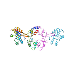
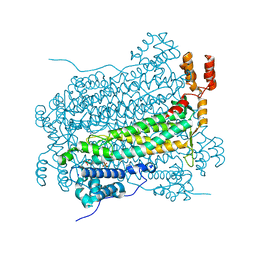
Crystal structure of phosphate-bound adenylosuccinate lyase from E. coli
Descriptor: Adenylosuccinate lyase, PHOSPHATE ION, SODIUM ION
Authors:Kozlov, G, Gehring, K, Montreal-Kingston Bacterial Structural Genomics Initiative (BSGI)
Deposit date:2009-04-07
Release date:2009-08-25
Last modified:2023-09-06
Method:X-RAY DIFFRACTION (1.9 Å)
Cite:The structure of phosphate-bound Escherichia coli adenylosuccinate lyase identifies His171 as a catalytic acid.
Acta Crystallogr.,Sect.F, 65, 2009

219140

PDB entries from 2024-05-01

PDB statisticsPDBj update infoContact PDBjnumon