Loading
PDBj
MenuPDBj@FacebookPDBj@TwitterPDBj@YouTubewwPDB FoundationwwPDB
RCSB PDBPDBeBMRBAdv. SearchSearch help
Search by PDB author
4R03
DownloadVisualize
BU of 4r03 by Molmil
Crystal structure of a DUF3836 family protein (BDI_3222) from Parabacteroides distasonis ATCC 8503 at 1.50 A resolution
Descriptor: (4S)-2-METHYL-2,4-PENTANEDIOL, CITRIC ACID, PHOSPHATE ION, ...
Authors:Joint Center for Structural Genomics (JCSG)
Deposit date:2014-07-29
Release date:2014-08-27
Last modified:2023-02-01
Method:X-RAY DIFFRACTION (1.5 Å)
Cite:Structures of single-layer beta-sheet proteins evolved from beta-hairpin repeats.
Protein Sci., 28, 2019
4RBO
DownloadVisualize
BU of 4rbo by Molmil
Crystal structure of a Nanog homeobox (NANOG) from Homo sapiens at 3.30 A resolution
Descriptor: 5'-D(*CP*TP*TP*GP*AP*AP*TP*GP*GP*GP*CP*C)-3', 5'-D(*GP*GP*CP*CP*CP*AP*TP*TP*CP*AP*AP*G)-3', Putative homeobox protein NANOGP8
Authors:Joint Center for Structural Genomics (JCSG), Partnership for Stem Cell Biology (STEMCELL)
Deposit date:2014-09-12
Release date:2014-10-01
Last modified:2023-02-01
Method:X-RAY DIFFRACTION (3.3 Å)
Cite:Structure-based discovery of NANOG variant with enhanced properties to promote self-renewal and reprogramming of pluripotent stem cells.
Proc.Natl.Acad.Sci.USA, 112, 2015
2RE3
DownloadVisualize
BU of 2re3 by Molmil
CRYSTAL STRUCTURE OF a DUF1285 family protein (SPO_0140) FROM SILICIBACTER POMEROYI DSS-3 AT 2.50 A RESOLUTION
Descriptor: GLYCEROL, Uncharacterized protein
Authors:Joint Center for Structural Genomics (JCSG)
Deposit date:2007-09-25
Release date:2007-10-09
Last modified:2023-01-25
Method:X-RAY DIFFRACTION (2.5 Å)
Cite:Structures of the first representatives of Pfam family PF06938 (DUF1285) reveal a new fold with repeated structural motifs and possible involvement in signal transduction.
Acta Crystallogr.,Sect.F, 66, 2010
1GAF
DownloadVisualize
BU of 1gaf by Molmil
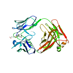
48G7 HYBRIDOMA LINE FAB COMPLEXED WITH HAPTEN 5-(PARA-NITROPHENYL PHOSPHONATE)-PENTANOIC ACID
Descriptor: 5-(PARA-NITROPHENYL PHOSPHONATE)-PENTANOIC ACID, CHIMERIC 48G7 FAB
Authors:Wedemayer, G.J, Patten, P.A, Stevens, R.C, Schultz, P.G.
Deposit date:1996-02-06
Release date:1996-07-11
Last modified:2011-07-13
Method:X-RAY DIFFRACTION (1.95 Å)
Cite:The immunological evolution of catalysis.
Science, 271, 1996
1HKL
DownloadVisualize
BU of 1hkl by Molmil
FREE AND LIGANDED FORM OF AN ESTEROLYTIC CATALYTIC ANTIBODY
Descriptor: 48G7 FAB
Authors:Wedemayer, G.J, Wang, L.H, Patten, P.A, Schultz, P.G, Stevens, R.C.
Deposit date:1996-12-20
Release date:1997-03-12
Last modified:2011-07-13
Method:X-RAY DIFFRACTION (2.68 Å)
Cite:Crystal structures of the free and liganded form of an esterolytic catalytic antibody.
J.Mol.Biol., 268, 1997
2PV7
DownloadVisualize
BU of 2pv7 by Molmil
Crystal structure of chorismate mutase / prephenate dehydrogenase (tyrA) (1574749) from Haemophilus influenzae RD at 2.00 A resolution
Descriptor: NICOTINAMIDE-ADENINE-DINUCLEOTIDE, T-protein [Includes: Chorismate mutase (EC 5.4.99.5) (CM) and Prephenate dehydrogenase (EC 1.3.1.12) (PDH)], TYROSINE
Authors:Joint Center for Structural Genomics (JCSG)
Deposit date:2007-05-09
Release date:2007-05-22
Last modified:2023-01-25
Method:X-RAY DIFFRACTION (2 Å)
Cite:The structure of Haemophilus influenzae prephenate dehydrogenase suggests unique features of bifunctional TyrA enzymes.
Acta Crystallogr.,Sect.F, 66, 2010
2Q3L
DownloadVisualize
BU of 2q3l by Molmil
CRYSTAL STRUCTURE OF AN UNCHARACTERIZED PROTEIN FROM DUF3478 FAMILY WITH A SPOIIAA-LIKE FOLD (SHEW_3102) FROM SHEWANELLA LOIHICA PV-4 AT 2.25 A RESOLUTION
Descriptor: (4S)-2-METHYL-2,4-PENTANEDIOL, CHLORIDE ION, SODIUM ION, ...
Authors:Joint Center for Structural Genomics (JCSG)
Deposit date:2007-05-30
Release date:2007-06-12
Last modified:2023-01-25
Method:X-RAY DIFFRACTION (2.25 Å)
Cite:Open and closed conformations of two SpoIIAA-like proteins (YP_749275.1 and YP_001095227.1) provide insights into membrane association and ligand binding.
Acta Crystallogr.,Sect.F, 66, 2010
2OOK
DownloadVisualize
BU of 2ook by Molmil
Crystal structure of a protein with unknown function (YP_749275.1) from Shewanella Frigidimarina NCIMB 400 at 1.80 A resolution
Descriptor: 1,2-ETHANEDIOL, Hypothetical protein
Authors:Joint Center for Structural Genomics (JCSG)
Deposit date:2007-01-25
Release date:2007-02-06
Last modified:2023-01-25
Method:X-RAY DIFFRACTION (1.8 Å)
Cite:Open and closed conformations of two SpoIIAA-like proteins (YP_749275.1 and YP_001095227.1) provide insights into membrane association and ligand binding.
Acta Crystallogr.,Sect.F, 66, 2010
2OOC
DownloadVisualize
BU of 2ooc by Molmil
Crystal structure of Histidine Phosphotransferase ShpA (NP_419930.1) from Caulobacter crescentus at 1.52 A resolution
Descriptor: GLYCEROL, Histidine phosphotransferase, TETRAETHYLENE GLYCOL
Authors:Joint Center for Structural Genomics (JCSG)
Deposit date:2007-01-25
Release date:2007-02-06
Last modified:2023-01-25
Method:X-RAY DIFFRACTION (1.52 Å)
Cite:Crystal structure of histidine phosphotransfer protein ShpA, an essential regulator of stalk biogenesis in Caulobacter crescentus.
J.Mol.Biol., 390, 2009
2Q8U
DownloadVisualize
BU of 2q8u by Molmil
CRYSTAL STRUCTURE OF MRE11 FROM THERMOTOGA MARITIMA MSB8 (TM1635) AT 2.20 A RESOLUTION
Descriptor: Exonuclease, putative
Authors:Joint Center for Structural Genomics (JCSG)
Deposit date:2007-06-11
Release date:2007-06-26
Last modified:2023-01-25
Method:X-RAY DIFFRACTION (2.2 Å)
Cite:Crystal structure of the first eubacterial Mre11 nuclease reveals novel features that may discriminate substrates during DNA repair.
J.Mol.Biol., 397, 2010
1AJ7
DownloadVisualize
BU of 1aj7 by Molmil
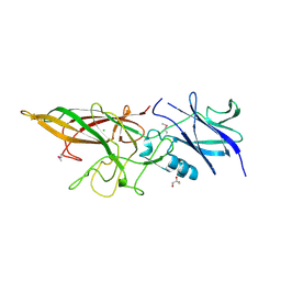
IMMUNOGLOBULIN 48G7 GERMLINE FAB ANTIBODY COMPLEXED WITH HAPTEN 5-(PARA-NITROPHENYL PHOSPHONATE)-PENTANOIC ACID. AFFINITY MATURATION OF AN ESTEROLYTIC ANTIBODY
Descriptor: 5-(PARA-NITROPHENYL PHOSPHONATE)-PENTANOIC ACID, IMMUNOGLOBULIN 48G7 FAB (HEAVY CHAIN), IMMUNOGLOBULIN 48G7 FAB (LIGHT CHAIN)
Authors:Wedemayer, G.J, Wang, L.H, Patten, P.A, Schultz, P.G, Stevens, R.C.
Deposit date:1997-05-15
Release date:1997-11-12
Last modified:2023-08-02
Method:X-RAY DIFFRACTION (2.1 Å)
Cite:Structural insights into the evolution of an antibody combining site.
Science, 276, 1997
2HUJ
DownloadVisualize
BU of 2huj by Molmil
Crystal structure of a protein of uknown function (NP_471338.1) from Listeria innocua at 1.74 A resolution
Descriptor: Lin2004 protein
Authors:Joint Center for Structural Genomics (JCSG)
Deposit date:2006-07-26
Release date:2006-08-08
Last modified:2023-01-25
Method:X-RAY DIFFRACTION (1.74 Å)
Cite:Crystal structures of MW1337R and lin2004: representatives of a novel protein family that adopt a four-helical bundle fold.
Proteins, 71, 2008
2IAY
DownloadVisualize
BU of 2iay by Molmil
Crystal structure of a duf1831 family protein (lp2179) from lactobacillus plantarum at 1.20 A resolution
Descriptor: CHLORIDE ION, GLYCEROL, Hypothetical protein
Authors:Joint Center for Structural Genomics (JCSG)
Deposit date:2006-09-08
Release date:2006-10-10
Last modified:2023-01-25
Method:X-RAY DIFFRACTION (1.2 Å)
Cite:Structure of LP2179, the first representative of Pfam family PF08866, suggests a new fold with a role in amino-acid metabolism.
Acta Crystallogr.,Sect.F, 66, 2010
2ICH
DownloadVisualize
BU of 2ich by Molmil
CRYSTAL STRUCTURE OF A PUTATIVE ATTH (NE1406) FROM NITROSOMONAS EUROPAEA AT 2.00 A RESOLUTION
Descriptor: 2-[N-CYCLOHEXYLAMINO]ETHANE SULFONIC ACID, GLYCEROL, Putative AttH, ...
Authors:Joint Center for Structural Genomics (JCSG)
Deposit date:2006-09-12
Release date:2006-10-03
Last modified:2023-01-25
Method:X-RAY DIFFRACTION (2 Å)
Cite:Structure of the first representative of Pfam family PF09410 (DUF2006) reveals a structural signature of the calycin superfamily that suggests a role in lipid metabolism.
Acta Crystallogr.,Sect.F, 66, 2010
2IIZ
DownloadVisualize
BU of 2iiz by Molmil
Crystal structure of putative melanin biosynthesis protein TyrA with bound heme (NP_716371.1) from Shewanella Oneidensis at 2.30 A resolution
Descriptor: 1,2-ETHANEDIOL, ISOPROPYL ALCOHOL, Melanin biosynthesis protein TyrA, ...
Authors:Joint Center for Structural Genomics (JCSG)
Deposit date:2006-09-28
Release date:2006-11-14
Last modified:2023-11-15
Method:X-RAY DIFFRACTION (2.3 Å)
Cite:Identification and structural characterization of heme binding in a novel dye-decolorizing peroxidase, TyrA.
Proteins, 69, 2007
2GHR
DownloadVisualize
BU of 2ghr by Molmil
Crystal structure of homoserine o-succinyltransferase (NP_981826.1) from Bacillus cereus ATCC 10987 at 2.40 A resolution
Descriptor: Homoserine O-succinyltransferase, SULFATE ION
Authors:Joint Center for Structural Genomics (JCSG)
Deposit date:2006-03-27
Release date:2006-04-11
Last modified:2023-01-25
Method:X-RAY DIFFRACTION (2.4 Å)
Cite:Crystal structure of homoserine O-succinyltransferase from Bacillus cereus at 2.4 A resolution
Proteins, 68, 2007
2GLZ
DownloadVisualize
BU of 2glz by Molmil
Crystal structure of a formylmethanofuran dehydrogenase subunit e-like protein (dhaf_2992) from desulfitobacterium hafniense dcb-2 at 1.45 A resolution
Descriptor: 1,2-ETHANEDIOL, NICKEL (II) ION, ZINC ION, ...
Authors:Joint Center for Structural Genomics (JCSG)
Deposit date:2006-04-05
Release date:2006-04-18
Last modified:2023-01-25
Method:X-RAY DIFFRACTION (1.45 Å)
Cite:Structures of three members of Pfam PF02663 (FmdE) implicated in microbial methanogenesis reveal a conserved alpha+beta core domain and an auxiliary C-terminal treble-clef zinc finger.
Acta Crystallogr.,Sect.F, 66, 2010
2GVI
DownloadVisualize
BU of 2gvi by Molmil
Crystal structure of a putative formylmethanofuran dehydrogenase subunit e (ta1109) from thermoplasma acidophilum at 1.87 A resolution
Descriptor: 1,2-ETHANEDIOL, ACETIC ACID, UNKNOWN LIGAND, ...
Authors:Joint Center for Structural Genomics (JCSG)
Deposit date:2006-05-02
Release date:2006-06-13
Last modified:2023-01-25
Method:X-RAY DIFFRACTION (1.87 Å)
Cite:Structures of three members of Pfam PF02663 (FmdE) implicated in microbial methanogenesis reveal a conserved alpha+beta core domain and an auxiliary C-terminal treble-clef zinc finger.
Acta Crystallogr.,Sect.F, 66, 2010
2HAG
DownloadVisualize
BU of 2hag by Molmil
Crystal structure of a putative dyp-type peroxidase protein (so_0740) from shewanella oneidensis at 2.75 A resolution
Descriptor: Melanin biosynthesis protein TyrA, putative
Authors:Joint Center for Structural Genomics (JCSG)
Deposit date:2006-06-12
Release date:2006-08-08
Last modified:2023-01-25
Method:X-RAY DIFFRACTION (2.75 Å)
Cite:Crystal structures of two novel dye-decolorizing peroxidases reveal a beta-barrel fold with a conserved heme-binding motif.
Proteins, 69, 2007
2HBW
DownloadVisualize
BU of 2hbw by Molmil
Crystal structure of a putative endopeptidase (ava_3396) from anabaena variabilis atcc 29413 at 1.05 A resolution
Descriptor: ACETATE ION, NLP/P60 protein, UNKNOWN LIGAND
Authors:Joint Center for Structural Genomics (JCSG)
Deposit date:2006-06-14
Release date:2006-08-08
Last modified:2023-01-25
Method:X-RAY DIFFRACTION (1.05 Å)
Cite:Structural Basis of Murein Peptide Specificity of a gamma-D-Glutamyl-L-Diamino Acid Endopeptidase.
Structure, 17, 2009
2GVK
DownloadVisualize
BU of 2gvk by Molmil
Crystal structure of a dye-decolorizing peroxidase (DyP) from Bacteroides thetaiotaomicron VPI-5482 at 1.6 A resolution
Descriptor: 1,2-ETHANEDIOL, ACETIC ACID, CHLORIDE ION, ...
Authors:Joint Center for Structural Genomics (JCSG)
Deposit date:2006-05-02
Release date:2006-05-16
Last modified:2023-01-25
Method:X-RAY DIFFRACTION (1.6 Å)
Cite:Crystal structures of two novel dye-decolorizing peroxidases reveal a beta-barrel fold with a conserved heme-binding motif.
Proteins, 69, 2007
2H1T
DownloadVisualize
BU of 2h1t by Molmil
Crystal structure of a duf1089 family protein (pa1994) from pseudomonas aeruginosa at 1.80 A resolution
Descriptor: (4S)-2-METHYL-2,4-PENTANEDIOL, 1,2-ETHANEDIOL, Hypothetical protein
Authors:Joint Center for Structural Genomics (JCSG)
Deposit date:2006-05-16
Release date:2006-05-30
Last modified:2023-01-25
Method:X-RAY DIFFRACTION (1.8 Å)
Cite:The structure of the first representative of Pfam family PF06475 reveals a new fold with possible involvement in glycolipid metabolism.
Acta Crystallogr.,Sect.F, 66, 2010
3LCT
DownloadVisualize
BU of 3lct by Molmil
Crystal Structure of the Anaplastic Lymphoma Kinase Catalytic Domain
Descriptor: ADENOSINE-5'-DIPHOSPHATE, ALK tyrosine kinase receptor, GLYCEROL
Authors:Lee, C.C.
Deposit date:2010-01-11
Release date:2010-08-04
Last modified:2023-09-06
Method:X-RAY DIFFRACTION (2.1 Å)
Cite:Crystal structure of the ALK (anaplastic lymphoma kinase) catalytic domain.
Biochem.J., 430, 2010
3L5O
DownloadVisualize
BU of 3l5o by Molmil
Crystal structure of protein with unknown function from DUF364 family (ZP_00559375.1) from Desulfitobacterium hafniense DCB-2 at 2.01 A resolution
Descriptor: 1,2-ETHANEDIOL, CHLORIDE ION, IMIDAZOLE, ...
Authors:Joint Center for Structural Genomics, Joint Center for Structural Genomics (JCSG)
Deposit date:2009-12-22
Release date:2010-02-02
Last modified:2023-02-01
Method:X-RAY DIFFRACTION (2.01 Å)
Cite:Structure of the first representative of Pfam family PF04016 (DUF364) reveals enolase and Rossmann-like folds that combine to form a unique active site with a possible role in heavy-metal chelation.
Acta Crystallogr.,Sect.F, 66, 2010
3LIU
DownloadVisualize
BU of 3liu by Molmil
Crystal structure of Putative cell adhesion protein (YP_001304840.1) from Parabacteroides distasonis ATCC 8503 at 2.05 A resolution
Descriptor: 1,2-ETHANEDIOL, CHLORIDE ION, GLYCEROL, ...
Authors:Joint Center for Structural Genomics (JCSG)
Deposit date:2010-01-25
Release date:2010-02-09
Last modified:2023-02-01
Method:X-RAY DIFFRACTION (2.05 Å)
Cite:A Distinct Type of Pilus from the Human Microbiome.
Cell, 165, 2016

221051

PDB entries from 2024-06-12

PDB statisticsPDBj update infoContact PDBjnumon