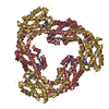
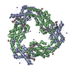
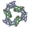
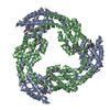
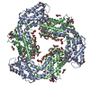
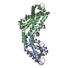
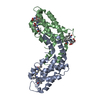
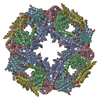
Journal: Acta Crystallogr D Biol Crystallogr / Year: 2014 Title: The structure of allophycocyanin B from Synechocystis PCC 6803 reveals the structural basis for the extreme redshift of the terminal emitter in phycobilisomes. Authors: Pan Pan Peng / Liang Liang Dong / Ya Fang Sun / Xiao Li Zeng / Wen Long Ding / Hugo Scheer / Xiaojing Yang / Kai Hong Zhao / Abstract: Allophycocyanin B (AP-B) is one of the two terminal emitters in phycobilisomes, the unique light-harvesting complexes of cyanobacteria and red algae. Its low excitation-energy level and the ...Allophycocyanin B (AP-B) is one of the two terminal emitters in phycobilisomes, the unique light-harvesting complexes of cyanobacteria and red algae. Its low excitation-energy level and the correspondingly redshifted absorption and fluorescence emission play an important role in funnelling excitation energy from the hundreds of chromophores of the extramembraneous phycobilisome to the reaction centres within the photosynthetic membrane. In the absence of crystal structures of these low-abundance terminal emitters, the molecular basis for the extreme redshift and directional energy transfer is largely unknown. Here, the crystal structure of trimeric AP-B [(ApcD/ApcB)3] from Synechocystis sp. PCC 6803 at 1.75 Å resolution is reported. In the crystal lattice, eight trimers of AP-B form a porous, spherical, 48-subunit assembly of 193 Å in diameter with an internal cavity of 1.1 × 10(6) Å(3). While the overall structure of trimeric AP-B is similar to those reported for many other phycobiliprotein trimers, the chromophore pocket of the α-subunit, ApcD, has more bulky residues that tightly pack the phycocyanobilin (PCB). Ring D of the chromophores is further stabilized by close interactions with ApcB from the adjacent monomer. The combined contributions from both subunits render the conjugated rings B, C and D of the PCB in ApcD almost perfectly coplanar. Together with mutagenesis data, it is proposed that the enhanced planarity effectively extends the conjugation system of PCB and leads to the redshifted absorption (λmax = 669 nm) and fluorescence emission (679 nm) of the ApcD chromophore in AP-B, thereby enabling highly efficient energy transfer from the phycobilisome core to the reaction centres.
In the structure databanks used in Yorodumi, some data are registered as the other names, "COVID-19 virus" and "2019-nCoV". Here are the details of the virus and the list of structure data.
Jan 31, 2019. EMDB accession codes are about to change! (news from PDBe EMDB page)
EMDB accession codes are about to change! (news from PDBe EMDB page)
The allocation of 4 digits for EMDB accession codes will soon come to an end. Whilst these codes will remain in use, new EMDB accession codes will include an additional digit and will expand incrementally as the available range of codes is exhausted. The current 4-digit format prefixed with “EMD-” (i.e. EMD-XXXX) will advance to a 5-digit format (i.e. EMD-XXXXX), and so on. It is currently estimated that the 4-digit codes will be depleted around Spring 2019, at which point the 5-digit format will come into force.
The EM Navigator/Yorodumi systems omit the EMD- prefix.
Related info.:Q: What is EMD? / ID/Accession-code notation in Yorodumi/EM Navigator
Yorodumi is a browser for structure data from EMDB, PDB, SASBDB, etc.
This page is also the successor to EM Navigator detail page, and also detail information page/front-end page for Omokage search.
The word "yorodu" (or yorozu) is an old Japanese word meaning "ten thousand". "mi" (miru) is to see.
Related info.:EMDB / PDB / SASBDB / Comparison of 3 databanks / Yorodumi Search / Aug 31, 2016. New EM Navigator & Yorodumi / Yorodumi Papers / Jmol/JSmol / Function and homology information / Changes in new EM Navigator and Yorodumi
 Movie
Movie Controller

 Yorodumi
 Open data
 Basic information
 Components
 Keywords
 PHOTOSYNTHESIS / alpha-helical phycobiliprotein / light harvesting /
  Function and homology information
 Authors
 Citation


 Structure visualization
 Downloads & links
 Other downloads
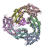


 PDBj
PDBj Assembly
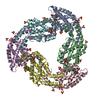

 Mass: 588.694 Da / Num. of mol.: 6 / Source method: obtained synthetically / Formula: C33H40N4O6
 Mass: 96.063 Da / Num. of mol.: 21 / Source method: obtained synthetically / Formula: SO4
 Mass: 18.015 Da / Num. of mol.: 1198 / Source method: isolated from a natural source / Formula: H2O
 Sample preparation
 Processing