 Movie
Movie Controller


+ Open data

- Basic information
| Entry | Database: PDB / ID: 5ook | ||||||
|---|---|---|---|---|---|---|---|
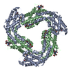
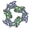
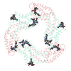
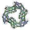
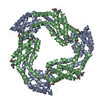
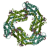
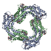
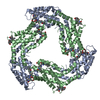
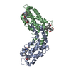
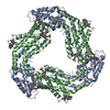
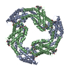
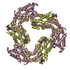
| Title | Structure of A. marina Phycocyanin contains overlapping isoforms | ||||||
 Components Components |
| ||||||
 Keywords Keywords |  PHOTOSYNTHESIS / Models / Phycobilisome / Phycocyanin / A. marina PHOTOSYNTHESIS / Models / Phycobilisome / Phycocyanin / A. marina | ||||||
| Function / homology |  Function and homology information: / phycobilisome / plasma membrane-derived thylakoid membrane / photosynthesis Function and homology information: / phycobilisome / plasma membrane-derived thylakoid membrane / photosynthesisSimilarity search - Function | ||||||
| Biological species |  Acaryochloris marina (bacteria) Acaryochloris marina (bacteria) | ||||||
| Method | X-RAY DIFFRACTION / SYNCHROTRON / MOLECULAR REPLACEMENT / Resolution: 2.1 Å | ||||||
 Authors Authors | Bar-Zvi, S. / Lahav, A. / Blankenship, E.R. / Adir, N. | ||||||
| Funding support |  Israel, 1items Israel, 1items
| ||||||
 Citation Citation | Journal: Biochim. Biophys. Acta / Year: 2018 Title: Structural heterogeneity leads to functional homogeneity in A. marina phycocyanin. Authors: Bar-Zvi, S. / Lahav, A. / Harris, D. / Niedzwiedzki, D.M. / Blankenship, R.E. / Adir, N. | ||||||
| History |
|
- Structure visualization
| Structure viewer | Molecule: MolmilJmol/JSmol |
|---|
- Downloads & links
-Download
| PDBx/mmCIF format | 5ook.cif.gz | 145.5 KB | Display | PDBx/mmCIF format |
|---|---|---|---|---|
| PDB format | pdb5ook.ent.gz | 116.4 KB | Display | PDB format |
| PDBx/mmJSON format | 5ook.json.gz | Tree view | PDBx/mmJSON format | |
| Others |  Other downloads Other downloads |
-Validation report
| Arichive directory | https://data.pdbj.org/pub/pdb/validation_reports/oo/5ookftp://data.pdbj.org/pub/pdb/validation_reports/oo/5ook | HTTPS FTP |
|---|
-Related structure data
| Related structure data | 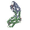 1cpcS S: Starting model for refinement |
|---|---|
| Similar structure data |
-Links
 PDBj
PDBj- Assembly
| Deposited unit | 
| ||||||||
|---|---|---|---|---|---|---|---|---|---|
| 1 | 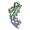
| ||||||||
| Unit cell |
|
-Components
| #1: Protein | Mass: 17390.361 Da / Num. of mol.: 1 / Source method: isolated from a natural source / Source: (natural) Acaryochloris marina (bacteria) / References: UniProt: A8ZMJ4 | ||||
|---|---|---|---|---|---|
| #2: Protein | Mass: 18038.377 Da / Num. of mol.: 1 / Source method: isolated from a natural source / Source: (natural) Acaryochloris marina (bacteria) / References: UniProt: A8ZMJ5 | ||||
| #3: Chemical | Phycocyanobilin  Mass: 588.694 Da / Num. of mol.: 3 / Source method: obtained synthetically / Formula: C33H40N4O6 / Feature type: SUBJECT OF INVESTIGATION Mass: 588.694 Da / Num. of mol.: 3 / Source method: obtained synthetically / Formula: C33H40N4O6 / Feature type: SUBJECT OF INVESTIGATION#4: Chemical | ChemComp-PEG / Diethylene glycol  Mass: 106.120 Da / Num. of mol.: 10 / Source method: obtained synthetically / Formula: C4H10O3 Mass: 106.120 Da / Num. of mol.: 10 / Source method: obtained synthetically / Formula: C4H10O3#5: Water | ChemComp-HOH / | Water Mass: 18.015 Da / Num. of mol.: 55 / Source method: isolated from a natural source / Formula: H2O Mass: 18.015 Da / Num. of mol.: 55 / Source method: isolated from a natural source / Formula: H2O |
-Experimental details
-Experiment
| Experiment | Method: X-RAY DIFFRACTION / Number of used crystals: 1 |
|---|
- Sample preparation
| Crystal | Density Matthews: 3.31 Å3/Da / Density % sol: 62.9 % |
|---|---|
| Crystal grow | Temperature: 293 K / Method: vapor diffusion, hanging drop / pH: 6.5 / Details: 0.1M HEPES pH 6.5, 0.1M MgCl2, 9% PEG 2K |
-Data collection
| Diffraction | Mean temperature: 100 K / Ambient temp details: 100 |
|---|---|
| Diffraction source | Source: SYNCHROTRON / Site: ESRF  / Beamline: ID29 / Wavelength: 0.976 Å / Beamline: ID29 / Wavelength: 0.976 Å |
| Detector | Type: DECTRIS PILATUS3 S 6M / Detector: PIXEL / Date: Nov 30, 2015 |
| Radiation | Protocol: SINGLE WAVELENGTH / Monochromatic (M) / Laue (L): M / Scattering type: x-ray |
| Radiation wavelength | Wavelength: 0.976 Å / Relative weight: 1 |
| Reflection | Resolution: 2.1→132.33 Å / Num. obs: 31134 / % possible obs: 99.9 % / Redundancy: 9.7 % / Biso Wilson estimate: 38.39 Å2 / CC1/2: 0.999 / Rmerge(I) obs: 0.069 / Rpim(I) all: 0.033 / Net I/σ(I): 10.6 |
| Reflection shell | Resolution: 2.1→2.175 Å / Redundancy: 10.5 % / Rmerge(I) obs: 0.555 / Mean I/σ(I) obs: 2.7 / Num. unique obs: 3105 / CC1/2: 0.92 / Rpim(I) all: 0.255 / % possible all: 99.9 |
- Processing
| Software |
| ||||||||||||||||||||||||||||||||||||||||||||||||||||||||||||||||||||||||||||||||||||||||||||||||||||||||||||||||||||||||||||||||||||||||||||||||||||||||||||||||||||||||||||||||||||||
|---|---|---|---|---|---|---|---|---|---|---|---|---|---|---|---|---|---|---|---|---|---|---|---|---|---|---|---|---|---|---|---|---|---|---|---|---|---|---|---|---|---|---|---|---|---|---|---|---|---|---|---|---|---|---|---|---|---|---|---|---|---|---|---|---|---|---|---|---|---|---|---|---|---|---|---|---|---|---|---|---|---|---|---|---|---|---|---|---|---|---|---|---|---|---|---|---|---|---|---|---|---|---|---|---|---|---|---|---|---|---|---|---|---|---|---|---|---|---|---|---|---|---|---|---|---|---|---|---|---|---|---|---|---|---|---|---|---|---|---|---|---|---|---|---|---|---|---|---|---|---|---|---|---|---|---|---|---|---|---|---|---|---|---|---|---|---|---|---|---|---|---|---|---|---|---|---|---|---|---|---|---|---|---|
| Refinement | Method to determine structure: MOLECULAR REPLACEMENT Starting model: 1CPC Resolution: 2.1→132.33 Å / Cor.coef. Fo:Fc: 0.963 / Cor.coef. Fo:Fc free: 0.953 / SU B: 9.551 / SU ML: 0.127 / Cross valid method: THROUGHOUT / ESU R: 0.159 / ESU R Free: 0.14 / Details: HYDROGENS HAVE BEEN ADDED IN THE RIDING POSITIONS
| ||||||||||||||||||||||||||||||||||||||||||||||||||||||||||||||||||||||||||||||||||||||||||||||||||||||||||||||||||||||||||||||||||||||||||||||||||||||||||||||||||||||||||||||||||||||
| Solvent computation | Ion probe radii: 0.9 Å / Shrinkage radii: 0.9 Å / VDW probe radii: 1.1 Å | ||||||||||||||||||||||||||||||||||||||||||||||||||||||||||||||||||||||||||||||||||||||||||||||||||||||||||||||||||||||||||||||||||||||||||||||||||||||||||||||||||||||||||||||||||||||
| Displacement parameters | Biso mean: 59.807 Å2
| ||||||||||||||||||||||||||||||||||||||||||||||||||||||||||||||||||||||||||||||||||||||||||||||||||||||||||||||||||||||||||||||||||||||||||||||||||||||||||||||||||||||||||||||||||||||
| Refinement step | Cycle: 1 / Resolution: 2.1→132.33 Å
| ||||||||||||||||||||||||||||||||||||||||||||||||||||||||||||||||||||||||||||||||||||||||||||||||||||||||||||||||||||||||||||||||||||||||||||||||||||||||||||||||||||||||||||||||||||||
| Refine LS restraints |
|