 Movie
Movie Controller

[English] 日本語
 Yorodumi
- PDB-3o2c: Crystal structure of a rod form of c-phycocyanin from Themosynech... -
+ Open data

- Basic information
| Entry | Database: PDB / ID: 3o2c | ||||||
|---|---|---|---|---|---|---|---|
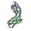
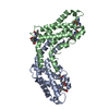
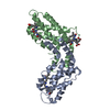
| Title | Crystal structure of a rod form of c-phycocyanin from Themosynechococcus vulcanus at 1.5 angstroms | ||||||
 Components Components |
| ||||||
 Keywords Keywords |  PHOTOSYNTHESIS / Phycobilisome / Cyanobacteria / Light Harvesting PHOTOSYNTHESIS / Phycobilisome / Cyanobacteria / Light Harvesting | ||||||
| Function / homology |  Function and homology informationphycobilisome / plasma membrane-derived thylakoid membrane / photosynthesis Function and homology informationphycobilisome / plasma membrane-derived thylakoid membrane / photosynthesisSimilarity search - Function | ||||||
| Biological species |  Thermosynechococcus vulcanus (bacteria) Thermosynechococcus vulcanus (bacteria) | ||||||
| Method | X-RAY DIFFRACTION / SYNCHROTRON / MOLECULAR REPLACEMENT / molecular replacement / Resolution: 1.5 Å | ||||||
 Authors Authors | David, L. / Marx, A. / Adir, N. | ||||||
 Citation Citation | Journal: J.Mol.Biol. / Year: 2011 Title: High-resolution crystal structures of trimeric and rod phycocyanin. Authors: David, L. / Marx, A. / Adir, N. | ||||||
| History |
|
- Structure visualization
| Structure viewer | Molecule: MolmilJmol/JSmol |
|---|
- Downloads & links
-Download
| PDBx/mmCIF format | 3o2c.cif.gz | 85 KB | Display | PDBx/mmCIF format |
|---|---|---|---|---|
| PDB format | pdb3o2c.ent.gz | 68.3 KB | Display | PDB format |
| PDBx/mmJSON format | 3o2c.json.gz | Tree view | PDBx/mmJSON format | |
| Others |  Other downloads Other downloads |
-Validation report
| Arichive directory | https://data.pdbj.org/pub/pdb/validation_reports/o2/3o2cftp://data.pdbj.org/pub/pdb/validation_reports/o2/3o2c | HTTPS FTP |
|---|
-Related structure data

-Links
 PDBj
PDBj- Assembly
| Deposited unit | 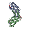
| ||||||||
|---|---|---|---|---|---|---|---|---|---|
| 1 | 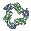
| ||||||||
| Unit cell |
| ||||||||
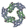
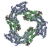
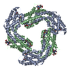
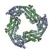
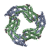
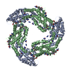
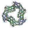
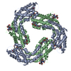
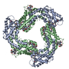
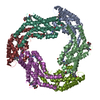
| Details | Authors state that phycocyanin forms hexamers which elongate into rod structures, a subcomponent of a large complex called the phycobilisome. |
-Components
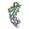
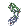
| #1: Protein | Mass: 17470.656 Da / Num. of mol.: 1 / Source method: isolated from a natural source / Source: (natural) Thermosynechococcus vulcanus (bacteria) / References: UniProt: Q9AM02 | ||
|---|---|---|---|
| #2: Protein | Mass: 18216.652 Da / Num. of mol.: 1 / Source method: isolated from a natural source / Source: (natural) Thermosynechococcus vulcanus (bacteria) / References: UniProt: Q71RW8 | ||
| #3: Chemical | Phycocyanobilin  Mass: 588.694 Da / Num. of mol.: 3 / Source method: obtained synthetically / Formula: C33H40N4O6 Mass: 588.694 Da / Num. of mol.: 3 / Source method: obtained synthetically / Formula: C33H40N4O6#4: Water | ChemComp-HOH / | Water Mass: 18.015 Da / Num. of mol.: 459 / Source method: isolated from a natural source / Formula: H2O Mass: 18.015 Da / Num. of mol.: 459 / Source method: isolated from a natural source / Formula: H2O |
-Experimental details
-Experiment
| Experiment | Method: X-RAY DIFFRACTION / Number of used crystals: 1 |
|---|
- Sample preparation
| Crystal | Density Matthews: 2.85 Å3/Da / Density % sol: 56.86 % |
|---|---|
| Crystal grow | Temperature: 293 K / Method: vapor diffusion, sitting drop / pH: 7 Details: 1.2M phosphate, pH 7, VAPOR DIFFUSION, SITTING DROP, temperature 293K |
-Data collection
| Diffraction source | Source: SYNCHROTRON / Site: ESRF  / Beamline: ID14-1 / Wavelength: 0.9795 Å / Beamline: ID14-1 / Wavelength: 0.9795 Å | ||||||||||||||||||||||||||||||||||||||||||||||||||||||||||||||||||||||||||||||||||||||||
|---|---|---|---|---|---|---|---|---|---|---|---|---|---|---|---|---|---|---|---|---|---|---|---|---|---|---|---|---|---|---|---|---|---|---|---|---|---|---|---|---|---|---|---|---|---|---|---|---|---|---|---|---|---|---|---|---|---|---|---|---|---|---|---|---|---|---|---|---|---|---|---|---|---|---|---|---|---|---|---|---|---|---|---|---|---|---|---|---|---|
| Radiation | Protocol: SINGLE WAVELENGTH / Monochromatic (M) / Laue (L): M / Scattering type: x-ray | ||||||||||||||||||||||||||||||||||||||||||||||||||||||||||||||||||||||||||||||||||||||||
| Radiation wavelength | Wavelength: 0.9795 Å / Relative weight: 1 | ||||||||||||||||||||||||||||||||||||||||||||||||||||||||||||||||||||||||||||||||||||||||
| Reflection | Resolution: 1.5→93.735 Å / Num. all: 63473 / Num. obs: 63473 / % possible obs: 98.8 % / Redundancy: 3.8 % / Rsym value: 0.019 / Net I/σ(I): 71.9 | ||||||||||||||||||||||||||||||||||||||||||||||||||||||||||||||||||||||||||||||||||||||||
| Reflection shell |
|
-Phasing
| Phasing | Method: molecular replacement | |||||||||
|---|---|---|---|---|---|---|---|---|---|---|
| Phasing MR | Rfactor: 35.9 / Model details: Phaser MODE: MR_AUTO
|
- Processing
| Software |
| ||||||||||||||||||||||||
|---|---|---|---|---|---|---|---|---|---|---|---|---|---|---|---|---|---|---|---|---|---|---|---|---|---|
| Refinement | Method to determine structure: MOLECULAR REPLACEMENT / Resolution: 1.5→26 Å / Cor.coef. Fo:Fc: 0.946 / Cor.coef. Fo:Fc free: 0.934 / Occupancy max: 1 / Occupancy min: 0.5 / SU B: 0.001 / SU ML: 0 / Cross valid method: THROUGHOUT / σ(F): 0 / ESU R Free: 0.092 / Stereochemistry target values: MAXIMUM LIKELIHOOD Details: HYDROGENS HAVE BEEN ADDED IN THE RIDING POSITIONS U VALUES : REFINED INDIVIDUALLY
| ||||||||||||||||||||||||
| Solvent computation | Ion probe radii: 0.8 Å / Shrinkage radii: 0.8 Å / VDW probe radii: 1.4 Å / Solvent model: MASK | ||||||||||||||||||||||||
| Displacement parameters | Biso max: 73.4 Å2 / Biso mean: 20.6126 Å2 / Biso min: 7.92 Å2
| ||||||||||||||||||||||||
| Refine analyze | Luzzati coordinate error obs: 0.32 Å / Luzzati d res low obs: 5 Å / Luzzati sigma a obs: 0.38 Å | ||||||||||||||||||||||||
| Refinement step | Cycle: LAST / Resolution: 1.5→26 Å
| ||||||||||||||||||||||||
| Refine LS restraints |
| ||||||||||||||||||||||||
| LS refinement shell | Resolution: 1.5→1.539 Å / Total num. of bins used: 20
|
