 Movie
Movie Controller

[English] 日本語
 Yorodumi
- PDB-1f99: CRYSTAL STRUCTURE OF R-PHYCOCYANIN FROM POLYSIPHONIA AT 2.4 A RES... -
+ Open data

- Basic information
| Entry | Database: PDB / ID: 1f99 | ||||||
|---|---|---|---|---|---|---|---|
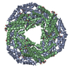
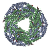
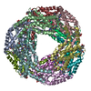
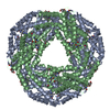
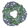
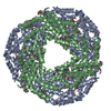
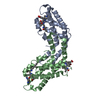
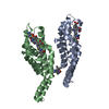
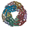
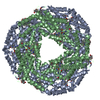
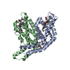
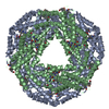
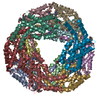
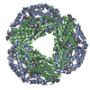
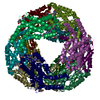
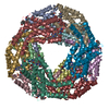
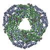
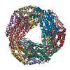
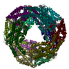
| Title | CRYSTAL STRUCTURE OF R-PHYCOCYANIN FROM POLYSIPHONIA AT 2.4 A RESOLUTION | ||||||
 Components Components | (R-PHYCOCYANIN) x 2 | ||||||
 Keywords Keywords |  PHOTOSYNTHESIS / LIGHT HARVESTING PROTEIN / R-PHYCOCYANIN / electron trasnport PHOTOSYNTHESIS / LIGHT HARVESTING PROTEIN / R-PHYCOCYANIN / electron trasnport | ||||||
| Function / homology |  Function and homology information Function and homology information | ||||||
| Biological species |  Polysiphonia urceolata (eukaryote) Polysiphonia urceolata (eukaryote) | ||||||
| Method | X-RAY DIFFRACTION / SYNCHROTRON / Resolution: 2.4 Å | ||||||
 Authors Authors | Liang, D.C. / Jiang, T. / Chang, W.R. | ||||||
 Citation Citation | Journal: Biophys.J. / Year: 2001 Title: Crystal structure of R-phycocyanin and possible energy transfer pathways in the phycobilisome. Authors: Jiang, T. / Zhang, J.P. / Chang, W.R. / Liang, D.C. | ||||||
| History |
|
- Structure visualization
| Structure viewer | Molecule: MolmilJmol/JSmol |
|---|
- Downloads & links
-Download
| PDBx/mmCIF format | 1f99.cif.gz | 203.9 KB | Display | PDBx/mmCIF format |
|---|---|---|---|---|
| PDB format | pdb1f99.ent.gz | 174.2 KB | Display | PDB format |
| PDBx/mmJSON format | 1f99.json.gz | Tree view | PDBx/mmJSON format | |
| Others |  Other downloads Other downloads |
-Validation report
| Arichive directory | https://data.pdbj.org/pub/pdb/validation_reports/f9/1f99ftp://data.pdbj.org/pub/pdb/validation_reports/f9/1f99 | HTTPS FTP |
|---|
-Related structure data
| Similar structure data |
|---|

-Links
 PDBj
PDBj- Assembly
| Deposited unit | 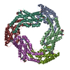
| ||||||||
|---|---|---|---|---|---|---|---|---|---|
| 1 | 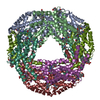
| ||||||||
| Unit cell |
| ||||||||
| Details | Bioloogical assembly is alpha 6 and beta 6, The asymmetry unit is alpha 3 and beta 3, |
-Components
-Protein , 2 types, 6 molecules AKMBLN
| #1: Protein | Mass: 17584.719 Da / Num. of mol.: 3 / Source method: isolated from a natural source / Details: alpha chain / Source: (natural) Polysiphonia urceolata (eukaryote) / References: UniProt: P59858#2: Protein | Mass: 18015.488 Da / Num. of mol.: 3 / Source method: isolated from a natural source / Details: beta chain / Source: (natural) Polysiphonia urceolata (eukaryote) / References: UniProt: P59859 |
|---|
-Non-polymers , 4 types, 349 molecules 


| #3: Chemical | ChemComp-BLA /  Mass: 582.646 Da / Num. of mol.: 1 / Source method: obtained synthetically / Formula: C33H34N4O6 Mass: 582.646 Da / Num. of mol.: 1 / Source method: obtained synthetically / Formula: C33H34N4O6 | ||||
|---|---|---|---|---|---|
| #4: Chemical | ChemComp-CYC / Phycocyanobilin Mass: 588.694 Da / Num. of mol.: 5 / Source method: obtained synthetically / Formula: C33H40N4O6 Mass: 588.694 Da / Num. of mol.: 5 / Source method: obtained synthetically / Formula: C33H40N4O6#5: Chemical | Phycoerythrobilin Mass: 588.694 Da / Num. of mol.: 3 / Source method: obtained synthetically / Formula: C33H40N4O6 Mass: 588.694 Da / Num. of mol.: 3 / Source method: obtained synthetically / Formula: C33H40N4O6#6: Water | ChemComp-HOH / | WaterMass: 18.015 Da / Num. of mol.: 340 / Source method: isolated from a natural source / Formula: H2O |
-Experimental details
-Experiment
| Experiment | Method: X-RAY DIFFRACTION / Number of used crystals: 1 |
|---|
- Sample preparation
| Crystal | Density Matthews: 4.48 Å3/Da / Density % sol: 40 % | ||||||||||||||||||||||||||||||||||||||||||
|---|---|---|---|---|---|---|---|---|---|---|---|---|---|---|---|---|---|---|---|---|---|---|---|---|---|---|---|---|---|---|---|---|---|---|---|---|---|---|---|---|---|---|---|
| Crystal grow | Temperature: 293 K / Method: vapor diffusion, hanging drop / pH: 7 Details: The hanging-drop contains 6mg/ml R-phycocyanin, 0.05M Na2HPO4/NaH2PO4 and (NH4)2SO4 (pH7.0). The diffusion buffer contained 10%-20% (NH4)2SO4 and 10% NaCl, 0.05M Na2HPO4/NaH2PO4, VAPOR DIFFUSION, HANGING DROP | ||||||||||||||||||||||||||||||||||||||||||
| Crystal grow | *PLUS pH: 7 | ||||||||||||||||||||||||||||||||||||||||||
| Components of the solutions | *PLUS
|
-Data collection
| Diffraction | Mean temperature: 277 K |
|---|---|
| Diffraction source | Source: SYNCHROTRON / Site: Photon Factory  / Beamline: BL-6B / Wavelength: 1 / Beamline: BL-6B / Wavelength: 1 |
| Detector | Type: WEISSENBERG / Detector: DIFFRACTOMETER / Date: Dec 10, 1997 |
| Radiation | Protocol: SINGLE WAVELENGTH / Monochromatic (M) / Laue (L): M / Scattering type: x-ray |
| Radiation wavelength | Wavelength: 1 Å / Relative weight: 1 |
| Reflection | Resolution: 2.4→20 Å / Num. all: 266953 / Num. obs: 73674 / % possible obs: 75.5 % / Observed criterion σ(F): 1 / Observed criterion σ(I): 1 / Redundancy: 3 % / Biso Wilson estimate: 16 Å2 / Rmerge(I) obs: 0.12 |
| Reflection shell | Resolution: 2.4→2.5 Å / Redundancy: 2.5 % / Rmerge(I) obs: 0.375 / % possible all: 91.1 |
| Reflection | *PLUS Num. measured all: 266953 |
| Reflection shell | *PLUS % possible obs: 49 % |
- Processing
| Software |
| ||||||||||||||||||||||||||||||||||||||||||||||||||||||||||||
|---|---|---|---|---|---|---|---|---|---|---|---|---|---|---|---|---|---|---|---|---|---|---|---|---|---|---|---|---|---|---|---|---|---|---|---|---|---|---|---|---|---|---|---|---|---|---|---|---|---|---|---|---|---|---|---|---|---|---|---|---|---|
| Refinement | Resolution: 2.4→8 Å / σ(F): 2 / σ(I): 2 / Stereochemistry target values: Engh & Huber
| ||||||||||||||||||||||||||||||||||||||||||||||||||||||||||||
| Refinement step | Cycle: LAST / Resolution: 2.4→8 Å
| ||||||||||||||||||||||||||||||||||||||||||||||||||||||||||||
| Refine LS restraints |
| ||||||||||||||||||||||||||||||||||||||||||||||||||||||||||||
| Software | *PLUS Name: X-PLOR / Classification: refinement | ||||||||||||||||||||||||||||||||||||||||||||||||||||||||||||
| Refinement | *PLUS Highest resolution: 2.4 Å / Lowest resolution: 8 Å / σ(F): 2 / % reflection Rfree: 10 % | ||||||||||||||||||||||||||||||||||||||||||||||||||||||||||||
| Solvent computation | *PLUS | ||||||||||||||||||||||||||||||||||||||||||||||||||||||||||||
| Displacement parameters | *PLUS | ||||||||||||||||||||||||||||||||||||||||||||||||||||||||||||
| Refine LS restraints | *PLUS
|
