 Movie
Movie Controller

[English] 日本語
 Yorodumi
- PDB-3rpw: The crystal structure of an ABC transporter from Rhodopseudomonas... -
+ Open data

- Basic information
| Entry | Database: PDB / ID: 3rpw | ||||||
|---|---|---|---|---|---|---|---|
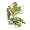
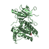
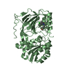
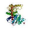
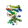
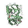
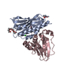
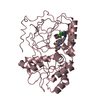
| Title | The crystal structure of an ABC transporter from Rhodopseudomonas palustris CGA009 | ||||||
 Components Components | ABC transporter ATP-binding cassette transporter ATP-binding cassette transporter | ||||||
 Keywords Keywords | TRANSPORT PROTEIN / structural genomics / PSI-Biology / protein structure initiative / midwest center for structural genomics / MCSG | ||||||
| Function / homology |  Function and homology information Function and homology information | ||||||
| Biological species |  Rhodopseudomonas palustris (phototrophic) Rhodopseudomonas palustris (phototrophic) | ||||||
| Method | X-RAY DIFFRACTION / SYNCHROTRON / SAD / Resolution: 1.65 Å | ||||||
 Authors Authors | Tan, K. / Mack, J.C. / Zerbs, S. / Collart, F. / Joachimiak, A. / Midwest Center for Structural Genomics (MCSG) | ||||||
 Citation Citation | Journal: Proteins / Year: 2013 Title: Structural and functional characterization of solute binding proteins for aromatic compounds derived from lignin: p-Coumaric acid and related aromatic acids. Authors: Tan, K. / Chang, C. / Cuff, M. / Osipiuk, J. / Landorf, E. / Mack, J.C. / Zerbs, S. / Joachimiak, A. / Collart, F.R. | ||||||
| History |
|
- Structure visualization
| Structure viewer | Molecule: MolmilJmol/JSmol |
|---|
- Downloads & links
-Download
| PDBx/mmCIF format | 3rpw.cif.gz | 152.5 KB | Display | PDBx/mmCIF format |
|---|---|---|---|---|
| PDB format | pdb3rpw.ent.gz | 126.9 KB | Display | PDB format |
| PDBx/mmJSON format | 3rpw.json.gz | Tree view | PDBx/mmJSON format | |
| Others |  Other downloads Other downloads |
-Validation report
| Arichive directory | https://data.pdbj.org/pub/pdb/validation_reports/rp/3rpwftp://data.pdbj.org/pub/pdb/validation_reports/rp/3rpw | HTTPS FTP |
|---|
-Related structure data
| Related structure data | 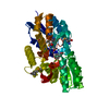 3sg0C 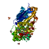 3tx6C  3uk0C 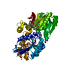 3ukjC  4dqdC 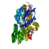 4eyoC 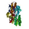 4eyqC 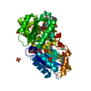 4f8jC 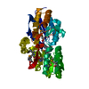 4fb4C 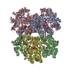 4i1dC C: citing same article ( |
|---|---|
| Similar structure data | |
| Other databases |

-Links
 PDBj
PDBj


- Assembly
| Deposited unit | 
| ||||||||
|---|---|---|---|---|---|---|---|---|---|
| 1 |
| ||||||||
| Unit cell |
| ||||||||
| Details | Experimentally unknown. It is predicted that the molecule is monomeric. |
-Components
| #1: Protein | ATP-binding cassette transporter Mass: 40730.086 Da / Num. of mol.: 1 Source method: isolated from a genetically manipulated source Source: (gene. exp.) Rhodopseudomonas palustris (phototrophic)Strain: CGA009 / Gene: RPA4648 / Plasmid: pMCSG7 / Production host: Escherichia coli (E. coli) / Strain (production host): BL21(DE3)magic / References: UniProt: Q6N0W2 | ||||||
|---|---|---|---|---|---|---|---|
| #2: Chemical | Glycerol  Mass: 92.094 Da / Num. of mol.: 3 / Source method: obtained synthetically / Formula: C3H8O3 Mass: 92.094 Da / Num. of mol.: 3 / Source method: obtained synthetically / Formula: C3H8O3#3: Chemical | ChemComp-FMT / Formic acid  Mass: 46.025 Da / Num. of mol.: 10 / Source method: obtained synthetically / Formula: CH2O2 Mass: 46.025 Da / Num. of mol.: 10 / Source method: obtained synthetically / Formula: CH2O2#4: Chemical | ChemComp-URE / | Urea  Mass: 60.055 Da / Num. of mol.: 1 / Source method: obtained synthetically / Formula: CH4N2O Mass: 60.055 Da / Num. of mol.: 1 / Source method: obtained synthetically / Formula: CH4N2O#5: Water | ChemComp-HOH / | Water Mass: 18.015 Da / Num. of mol.: 227 / Source method: isolated from a natural source / Formula: H2O Mass: 18.015 Da / Num. of mol.: 227 / Source method: isolated from a natural source / Formula: H2O |
-Experimental details
-Experiment
| Experiment | Method: X-RAY DIFFRACTION / Number of used crystals: 1 |
|---|
- Sample preparation
| Crystal | Density Matthews: 2.53 Å3/Da / Density % sol: 51.41 % |
|---|---|
| Crystal grow | Temperature: 289 K / Method: vapor diffusion, sitting drop / pH: 7 Details: 2.4M Sodium Malonate, pH 7.0, VAPOR DIFFUSION, SITTING DROP, temperature 289K |
-Data collection
| Diffraction | Mean temperature: 100 K |
|---|---|
| Diffraction source | Source: SYNCHROTRON / Site: APS  / Beamline: 19-BM / Wavelength: 0.97923 Å / Beamline: 19-BM / Wavelength: 0.97923 Å |
| Detector | Type: ADSC QUANTUM 315r / Detector: CCD / Date: Apr 22, 2011 / Details: mirror |
| Radiation | Monochromator: Si 111 crystal / Protocol: SINGLE WAVELENGTH / Monochromatic (M) / Laue (L): M / Scattering type: x-ray |
| Radiation wavelength | Wavelength: 0.97923 Å / Relative weight: 1 |
| Reflection | Resolution: 1.65→32.5 Å / Num. all: 50234 / Num. obs: 50234 / % possible obs: 99.8 % / Observed criterion σ(F): 0 / Observed criterion σ(I): 0 / Redundancy: 6 % / Rmerge(I) obs: 0.084 / Net I/σ(I): 36.3 |
| Reflection shell | Resolution: 1.65→1.68 Å / Redundancy: 5.9 % / Rmerge(I) obs: 0.617 / Mean I/σ(I) obs: 2.8 / Num. unique all: 2531 / % possible all: 100 |
- Processing
| Software |
| |||||||||||||||||||||||||||||||||||||||||||||||||||||||||||||||||||||||||||||||||||||||||||||||||||||||||||||||||||||||||||||||||||||
|---|---|---|---|---|---|---|---|---|---|---|---|---|---|---|---|---|---|---|---|---|---|---|---|---|---|---|---|---|---|---|---|---|---|---|---|---|---|---|---|---|---|---|---|---|---|---|---|---|---|---|---|---|---|---|---|---|---|---|---|---|---|---|---|---|---|---|---|---|---|---|---|---|---|---|---|---|---|---|---|---|---|---|---|---|---|---|---|---|---|---|---|---|---|---|---|---|---|---|---|---|---|---|---|---|---|---|---|---|---|---|---|---|---|---|---|---|---|---|---|---|---|---|---|---|---|---|---|---|---|---|---|---|---|---|
| Refinement | Method to determine structure: SAD / Resolution: 1.65→32.5 Å / SU ML: 0.17 / σ(F): 0 / Phase error: 17.88 / Stereochemistry target values: ML
| |||||||||||||||||||||||||||||||||||||||||||||||||||||||||||||||||||||||||||||||||||||||||||||||||||||||||||||||||||||||||||||||||||||
| Solvent computation | Shrinkage radii: 0.72 Å / VDW probe radii: 1 Å / Solvent model: FLAT BULK SOLVENT MODEL / Bsol: 63.898 Å2 / ksol: 0.438 e/Å3 | |||||||||||||||||||||||||||||||||||||||||||||||||||||||||||||||||||||||||||||||||||||||||||||||||||||||||||||||||||||||||||||||||||||
| Displacement parameters |
| |||||||||||||||||||||||||||||||||||||||||||||||||||||||||||||||||||||||||||||||||||||||||||||||||||||||||||||||||||||||||||||||||||||
| Refinement step | Cycle: LAST / Resolution: 1.65→32.5 Å
| |||||||||||||||||||||||||||||||||||||||||||||||||||||||||||||||||||||||||||||||||||||||||||||||||||||||||||||||||||||||||||||||||||||
| Refine LS restraints |
| |||||||||||||||||||||||||||||||||||||||||||||||||||||||||||||||||||||||||||||||||||||||||||||||||||||||||||||||||||||||||||||||||||||
| LS refinement shell |
| |||||||||||||||||||||||||||||||||||||||||||||||||||||||||||||||||||||||||||||||||||||||||||||||||||||||||||||||||||||||||||||||||||||
| Refinement TLS params. | Method: refined / Origin x: 3.0055 Å / Origin y: 50.4873 Å / Origin z: 20.4975 Å
| |||||||||||||||||||||||||||||||||||||||||||||||||||||||||||||||||||||||||||||||||||||||||||||||||||||||||||||||||||||||||||||||||||||
| Refinement TLS group | Selection details: all |
