 Movie
Movie Controller


+ Open data

- Basic information
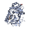
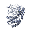
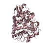
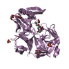
| Entry | Database: PDB / ID: 3vzb | ||||||
|---|---|---|---|---|---|---|---|
| Title | Crystal structure of Sphingosine Kinase 1 | ||||||
 Components Components | Sphingosine kinase 1 | ||||||
 Keywords Keywords | TRANSFERASE/INHIBITOR / lipid kinase / TRANSFERASE-INHIBITOR complex | ||||||
| Function / homology |  Function and homology informationsphingosine kinase / sphinganine kinase activity / D-erythro-sphingosine kinase activity / sphingoid catabolic process / regulation of endosomal vesicle fusion / sphingosine metabolic process / negative regulation of ceramide biosynthetic process / sphingosine-1-phosphate receptor activity / regulation of microglial cell activation / regulation of interleukin-1 beta production ...sphingosine kinase / sphinganine kinase activity / D-erythro-sphingosine kinase activity / sphingoid catabolic process / regulation of endosomal vesicle fusion / sphingosine metabolic process / negative regulation of ceramide biosynthetic process / sphingosine-1-phosphate receptor activity / regulation of microglial cell activation / regulation of interleukin-1 beta production / sphingosine biosynthetic process / sphingolipid biosynthetic process / regulation of neuroinflammatory response / Sphingolipid de novo biosynthesis / positive regulation of smooth muscle contraction / regulation of phagocytosis / regulation of tumor necrosis factor-mediated signaling pathway / positive regulation of p38MAPK cascade / DNA biosynthetic process / protein acetylation / blood vessel development / positive regulation of interleukin-17 production / Association of TriC/CCT with target proteins during biosynthesis / acetyltransferase activity / regulation of endocytosis / endocytic vesicle / cellular response to vascular endothelial growth factor stimulus / response to tumor necrosis factor / clathrin-coated pit / Transferases; Acyltransferases; Transferring groups other than aminoacyl groups / positive regulation of mitotic cell cycle / positive regulation of mitotic nuclear division / protein phosphatase 2A binding / positive regulation of peptidyl-threonine phosphorylation / VEGFR2 mediated cell proliferation / positive regulation of protein ubiquitination / calcium-mediated signaling / brain development / PKR-mediated signaling / cellular response to growth factor stimulus / cellular response to hydrogen peroxide / positive regulation of non-canonical NF-kappaB signal transduction / positive regulation of angiogenesis / positive regulation of fibroblast proliferation / presynapse / positive regulation of NF-kappaB transcription factor activity / early endosome membrane / positive regulation of cell growth / cell population proliferation / Extra-nuclear estrogen signaling / calmodulin binding / intracellular signal transduction / positive regulation of cell migration / inflammatory response / phosphorylation / intracellular membrane-bounded organelle / lipid binding / negative regulation of apoptotic process / magnesium ion binding / DNA binding / nucleoplasm / ATP binding / membrane / nucleus / plasma membrane / cytosol / cytoplasm Function and homology informationsphingosine kinase / sphinganine kinase activity / D-erythro-sphingosine kinase activity / sphingoid catabolic process / regulation of endosomal vesicle fusion / sphingosine metabolic process / negative regulation of ceramide biosynthetic process / sphingosine-1-phosphate receptor activity / regulation of microglial cell activation / regulation of interleukin-1 beta production ...sphingosine kinase / sphinganine kinase activity / D-erythro-sphingosine kinase activity / sphingoid catabolic process / regulation of endosomal vesicle fusion / sphingosine metabolic process / negative regulation of ceramide biosynthetic process / sphingosine-1-phosphate receptor activity / regulation of microglial cell activation / regulation of interleukin-1 beta production / sphingosine biosynthetic process / sphingolipid biosynthetic process / regulation of neuroinflammatory response / Sphingolipid de novo biosynthesis / positive regulation of smooth muscle contraction / regulation of phagocytosis / regulation of tumor necrosis factor-mediated signaling pathway / positive regulation of p38MAPK cascade / DNA biosynthetic process / protein acetylation / blood vessel development / positive regulation of interleukin-17 production / Association of TriC/CCT with target proteins during biosynthesis / acetyltransferase activity / regulation of endocytosis / endocytic vesicle / cellular response to vascular endothelial growth factor stimulus / response to tumor necrosis factor / clathrin-coated pit / Transferases; Acyltransferases; Transferring groups other than aminoacyl groups / positive regulation of mitotic cell cycle / positive regulation of mitotic nuclear division / protein phosphatase 2A binding / positive regulation of peptidyl-threonine phosphorylation / VEGFR2 mediated cell proliferation / positive regulation of protein ubiquitination / calcium-mediated signaling / brain development / PKR-mediated signaling / cellular response to growth factor stimulus / cellular response to hydrogen peroxide / positive regulation of non-canonical NF-kappaB signal transduction / positive regulation of angiogenesis / positive regulation of fibroblast proliferation / presynapse / positive regulation of NF-kappaB transcription factor activity / early endosome membrane / positive regulation of cell growth / cell population proliferation / Extra-nuclear estrogen signaling / calmodulin binding / intracellular signal transduction / positive regulation of cell migration / inflammatory response / phosphorylation / intracellular membrane-bounded organelle / lipid binding / negative regulation of apoptotic process / magnesium ion binding / DNA binding / nucleoplasm / ATP binding / membrane / nucleus / plasma membrane / cytosol / cytoplasmSimilarity search - Function | ||||||
| Biological species |  Homo sapiens (human) Homo sapiens (human) | ||||||
| Method | X-RAY DIFFRACTION / SYNCHROTRON / MAD / Resolution: 2 Å | ||||||
 Authors Authors | Min, X. / Walker, N.P. / Wang, Z. | ||||||
 Citation Citation | Journal: Structure / Year: 2013 Title: Molecular basis of sphingosine kinase 1 substrate recognition and catalysis. Authors: Wang, Z. / Min, X. / Xiao, S.H. / Johnstone, S. / Romanow, W. / Meininger, D. / Xu, H. / Liu, J. / Dai, J. / An, S. / Thibault, S. / Walker, N. | ||||||
| History |
|
- Structure visualization
| Structure viewer | Molecule: MolmilJmol/JSmol |
|---|
- Downloads & links
-Download
| PDBx/mmCIF format | 3vzb.cif.gz | 222 KB | Display | PDBx/mmCIF format |
|---|---|---|---|---|
| PDB format | pdb3vzb.ent.gz | 179.7 KB | Display | PDB format |
| PDBx/mmJSON format | 3vzb.json.gz | Tree view | PDBx/mmJSON format | |
| Others |  Other downloads Other downloads |
-Validation report
| Arichive directory | https://data.pdbj.org/pub/pdb/validation_reports/vz/3vzbftp://data.pdbj.org/pub/pdb/validation_reports/vz/3vzb | HTTPS FTP |
|---|
-Related structure data


-Links
 PDBj
PDBj


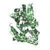
- Assembly
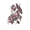
| Deposited unit | 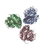
| ||||||||
|---|---|---|---|---|---|---|---|---|---|
| 1 | 
| ||||||||
| 2 | 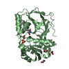
| ||||||||
| 3 | 
| ||||||||
| Unit cell |
|
-Components
| #1: Protein | / SK 1 / SPK 1 Mass: 39860.422 Da / Num. of mol.: 3 / Fragment: UNP RESIDUES 9-364 Source method: isolated from a genetically manipulated source Source: (gene. exp.) Homo sapiens (human) / Cell line: sf9 / Gene: SPHK1, SPHK, SPK / Plasmid: pFastBac HTb / References: UniProt: Q9NYA1, sphingosine kinase#2: Chemical | Sphingosine  Mass: 299.492 Da / Num. of mol.: 2 / Source method: obtained synthetically / Formula: C18H37NO2 Mass: 299.492 Da / Num. of mol.: 2 / Source method: obtained synthetically / Formula: C18H37NO2#3: Chemical | Sulfate  Mass: 96.063 Da / Num. of mol.: 3 / Source method: obtained synthetically / Formula: SO4 Mass: 96.063 Da / Num. of mol.: 3 / Source method: obtained synthetically / Formula: SO4#4: Chemical | ChemComp-EDO / Ethylene glycol  Mass: 62.068 Da / Num. of mol.: 15 / Source method: obtained synthetically / Formula: C2H6O2 Mass: 62.068 Da / Num. of mol.: 15 / Source method: obtained synthetically / Formula: C2H6O2#5: Water | ChemComp-HOH / | Water Mass: 18.015 Da / Num. of mol.: 384 / Source method: isolated from a natural source / Formula: H2O Mass: 18.015 Da / Num. of mol.: 384 / Source method: isolated from a natural source / Formula: H2O |
|---|
-Experimental details
-Experiment
| Experiment | Method: X-RAY DIFFRACTION / Number of used crystals: 1 |
|---|
- Sample preparation
| Crystal | Density Matthews: 2.57 Å3/Da / Density % sol: 52.05 % |
|---|---|
| Crystal grow | Temperature: 289 K / Method: sitting drop / pH: 6 Details: 1.0-1.4M ammonium sulfate, 0.3-1.3M NaCl, 0.1M Bis-Tris,, pH 6.0, sitting drop, temperature 289K |
-Data collection
| Diffraction | Mean temperature: 100 K | |||||||||||||||||||||||||||||||||||||||||||||||||||||||||||||||||||||||||||||
|---|---|---|---|---|---|---|---|---|---|---|---|---|---|---|---|---|---|---|---|---|---|---|---|---|---|---|---|---|---|---|---|---|---|---|---|---|---|---|---|---|---|---|---|---|---|---|---|---|---|---|---|---|---|---|---|---|---|---|---|---|---|---|---|---|---|---|---|---|---|---|---|---|---|---|---|---|---|---|
| Diffraction source | Source: SYNCHROTRON / Site: ALS  / Beamline: 5.0.2 / Wavelength: 1 Å / Beamline: 5.0.2 / Wavelength: 1 Å | |||||||||||||||||||||||||||||||||||||||||||||||||||||||||||||||||||||||||||||
| Detector | Type: ADSC315R / Detector: CCD / Details: 3x3 CCD array | |||||||||||||||||||||||||||||||||||||||||||||||||||||||||||||||||||||||||||||
| Radiation | Monochromator: Double-crystal, Si(111) / Protocol: SINGLE WAVELENGTH / Scattering type: x-ray | |||||||||||||||||||||||||||||||||||||||||||||||||||||||||||||||||||||||||||||
| Radiation wavelength | Wavelength: 1 Å / Relative weight: 1 | |||||||||||||||||||||||||||||||||||||||||||||||||||||||||||||||||||||||||||||
| Reflection | Resolution: 2→113.131 Å / Num. all: 82888 / Num. obs: 82888 / % possible obs: 99.7 % / Redundancy: 4.3 % / Rsym value: 0.062 / Net I/σ(I): 13.2 | |||||||||||||||||||||||||||||||||||||||||||||||||||||||||||||||||||||||||||||
| Reflection shell |
|
- Processing
| Software |
| ||||||||||||||||||||||||||||||||||||||||||||||||||||||||||||||||||||||||||||||||||||||||||||||||||||||||||||||||||||||||||||||||||||||||||||||||||||||||||||||||||||||||||
|---|---|---|---|---|---|---|---|---|---|---|---|---|---|---|---|---|---|---|---|---|---|---|---|---|---|---|---|---|---|---|---|---|---|---|---|---|---|---|---|---|---|---|---|---|---|---|---|---|---|---|---|---|---|---|---|---|---|---|---|---|---|---|---|---|---|---|---|---|---|---|---|---|---|---|---|---|---|---|---|---|---|---|---|---|---|---|---|---|---|---|---|---|---|---|---|---|---|---|---|---|---|---|---|---|---|---|---|---|---|---|---|---|---|---|---|---|---|---|---|---|---|---|---|---|---|---|---|---|---|---|---|---|---|---|---|---|---|---|---|---|---|---|---|---|---|---|---|---|---|---|---|---|---|---|---|---|---|---|---|---|---|---|---|---|---|---|---|---|---|---|---|
| Refinement | Method to determine structure: MAD / Resolution: 2→48.22 Å / Cor.coef. Fo:Fc: 0.951 / Cor.coef. Fo:Fc free: 0.925 / Occupancy max: 1 / Occupancy min: 0.5 / SU B: 4.647 / SU ML: 0.13 / SU R Cruickshank DPI: 0.1947 / Cross valid method: THROUGHOUT / ESU R: 0.192 / ESU R Free: 0.177 / Stereochemistry target values: MAXIMUM LIKELIHOOD / Details: HYDROGENS HAVE BEEN ADDED IN THE RIDING POSITIONS
| ||||||||||||||||||||||||||||||||||||||||||||||||||||||||||||||||||||||||||||||||||||||||||||||||||||||||||||||||||||||||||||||||||||||||||||||||||||||||||||||||||||||||||
| Solvent computation | Ion probe radii: 0.8 Å / Shrinkage radii: 0.8 Å / VDW probe radii: 1.2 Å / Solvent model: MASK | ||||||||||||||||||||||||||||||||||||||||||||||||||||||||||||||||||||||||||||||||||||||||||||||||||||||||||||||||||||||||||||||||||||||||||||||||||||||||||||||||||||||||||
| Displacement parameters | Biso mean: 39.712 Å2
| ||||||||||||||||||||||||||||||||||||||||||||||||||||||||||||||||||||||||||||||||||||||||||||||||||||||||||||||||||||||||||||||||||||||||||||||||||||||||||||||||||||||||||
| Refinement step | Cycle: LAST / Resolution: 2→48.22 Å
| ||||||||||||||||||||||||||||||||||||||||||||||||||||||||||||||||||||||||||||||||||||||||||||||||||||||||||||||||||||||||||||||||||||||||||||||||||||||||||||||||||||||||||
| Refine LS restraints |
| ||||||||||||||||||||||||||||||||||||||||||||||||||||||||||||||||||||||||||||||||||||||||||||||||||||||||||||||||||||||||||||||||||||||||||||||||||||||||||||||||||||||||||
| LS refinement shell | Resolution: 2→2.052 Å / Total num. of bins used: 20
|
